The cerebrospinal division is part of the nervous membranes of the Central type. It occupies the vertebral canal. The external structure of the dorsal plot be in the form of a very thick tube having tapered walls in the inner part, and the shell at both the front and rear.
The spinal division
The structure of the spinal cord is characterized by a very deep structure and the main function of the spinal cord is to transmit impulses from the brain to the peripheral nervous system type. This should be the procedure of the reflex sample.
The spinal cord of the child and the adult responsible for the natural breathing process, proper heart function, normal digestion, sexual activity, and also any bodily movement.
In this article you will have the opportunity to learn the following:
- What are the functions of spinal cord?
- What are the reflex arcs of the spinal cord?
- The psychophysiology the spinal activities.
- The structure and function of the brain and spinal cord – what are the differences?
- What is the feature of the structure of the spinal cord in young children (preschoolers)?
- Morphofunctional function of the spinal cord.
- What is included in the segmental apparatus of the spinal cord?
- Which element of the somatic reflex arc is located entirely in the spinal cord?
It is worth mentioning that the spinal area is usually created by the fourth week the baby’s development inside the womb. It happens in the period when the woman can not even suspect about the pregnancy. During pregnancy is differentiation of heterogeneous elements, certain spinal departments also finally formed the first two years of life.

We present our material on the subject: “Structure, functions and age-related features of the spinal cord”.
Features of the external structure
The initial separation of the body shall be determined conventionally on the edge of the first vertebra transverse division, as well as large hole in the skull in the neck. In this place the spinal part flows smoothly into the part of the brain. While the exact distinction between the brain and the spinal cord is simply no. This should be a kind of decussation of the pyramidal paths: paths that are responsible for motor function of the limbs. The spinal region, located below, will correspond to the upper part of the second lumbar vertebra.
For the prevention and treatment of diseases of the JOINTS our constant reader uses the increasingly popular NON-surgical method of treatment is recommended by leading German and Israeli orthopedists. Thoroughly acquainted with him, we decided to offer it to your attention.
Thus, the overall length of the body is much less than the length of the spinal canal. Specifically, this feature of localization of the spinal Department will provide an opportunity for the puncture of the dorsal part of the third and fourth vertebrae in the lumbar region (damage to the spinal area in the case of lumbar puncture of the sample between the spinous processes of the third and fourth vertebrae is almost impossible, because in this area they simply do not exist).
A person on the Central nervous system has the following dimensions: the total length of the body ranges from forty to forty-five centimeters, the thickness is from one to one and a half inches, and weighs from thirty to thirty-five grams.

In terms of length usually allocate a certain amount of cerebrospinal departments:
Found effective remedy for pains and for the treatment of joints:
- natural composition,
- with no side effects
- efficiency, proven expert,
- a quick result.
- cervical;
- lower back;
- the chest part;
- the sacrum;
- the coccyx.
In the lumbosacral and cervical levels of the spinal part would be thicker than in the other divisions, because in these places there is a maximum concentration of cells of the nervous type, which are responsible for limb movements.
End segments sacral coccygeal sample represent a cone area of the spinal cord, because is characterized by the corresponding geometrical shape of the sample. This taper provides a gradual transition in the filament terminal of the sample. This thread can contain only connective tissue, not having elements of the nerve cells. The fabrics are the spinal sheath. The terminal filament is attached to the second coccygeal vertebra.
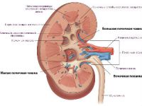
On the Central nervous system along the entire length is covered by three membranes of the brain nature. 1-I shell body is soft. It contains the venous and arterial vessels of the type that conduct the blood to all parts of the body. The second sheath is the middle, or arachnoid. The space between these 2 membranes is called the subarachnoid, it contains special fluid – the cerebrospinal fluid. When performing a spinal tap, the needle must be exactly in the given space to take the substance on a thorough analysis. The outer shell of the body has a solid, durable structure.
Sheath occupies a very large space, reaching to the holes between the vertebrae, passing parallel to the spine, which constitutes a part of the nervous system (your own machine spinal cord).
In the inner compartment of the vertebral canal area of the spinal cord is fixed on the outer shell of the vertebrae, using the cords. In the Central part of the body is a narrow tube constituting the main channel.
What is the neuron
Before you begin to discuss the functional significance of the spinal cord, it is necessary to tell that represent the neurons. The motoneurons of the spinal cord – the large cells of the nervous type, which occupy major parts of the brain spinal cord (morpho-functional organization of the spinal cord). These cells are responsible for motor coordination process and maintaining muscle tone.

Motoneurons, located on the periphery of the spinal cord are classified into two main types: gamma and alpha.
The motoneurons are, in fact, is a neural organization of the spinal cord. Neurons of the gamma type have a small size, they Innervate the fibers of the muscles intravazalnoe sample. Spinal reflexes are usually reduce with the help of alpha neurons.
Alpha/gamma neurons can be inhibited by reason of the action of neurons in the intercalated sample, and due to the fact that motor functions of the spinal cord show strong activity. The functional anatomy of the spinal cord and age-related features of the spinal cord directly dependent on the activity of motoneurons that operate in the field of autonomic nervous system type.
The structure and function of the spinal division
Finally, it was time to discuss what the spinal cord has a characteristic structure and functions. In the direction of the node element is branched at a special segmental structure (departments). Each individual segment has a branching anterior and posterior roots. The first responsible for the process of transfer of information related to movements due to what are considered to be motor. The roots of the back of the sample saturate the brain spinal information, i.e. give data related sensations due to what they called the sensitive.
The spinal cord performs the following functions:
- Conduction function of the spinal cord.
- Reflex function of the spinal cord.
A brief analysis of the features, you have the opportunity to evaluate the table:
| Functions of the spinal cord | |
| Reflex
Gray matter |
Conductor
White matter |
| Conducting pulse sites on the body muscles through a top-down conductive nodes. | Implementation of the sensitive pulse pushes from the skin, strong tendons, articular areas, of abnormal receptors and temperature. |
| Implementing major decisions. | On the basic ways, the relationship of the brain and spinal cord. |
Here you can also know which of these functions does not fulfill the spinal cord. Reflex and conductive functions are basic. Their activities affect directly the age peculiarities of the spinal cord. We will try to understand how the body and its functions, activate the circulatory system of the body, and what should be the physiological feature of the spinal segment.
The power of the Central organ of the nervous system is provided by the blood vessels of the type that are conducted from the aorta, and arterial compartments. The upper segments of the cervical fueled by blood coming from the arteries to the vertebral type (as in the case of the brain) through the spinal compartment.
The reflex centers of the spinal cord are responsible for the system response of an organism to external stimuli. Basically everything we do daily is associated with activity generated by spinal reflexes. The spinal reflexes are mainly responsible of the reflex arc. The reflex arc is responsible for the response to external irritation. Under this scenario attempts produced by a reflex arc, not controlled by the brain part of the head.
Conductor apparatus of the spinal cord responsible for the transmission of short pulses the peripheral area to the Central compartment. The spinal cord performs across the conductors of the transmission information data in all directions.