
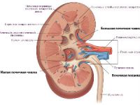
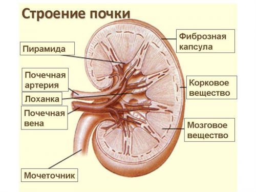
The anatomy of the kidney identify some features associated with the location of the pelvis, cups, pyramids, cavities and recesses. One of these recesses is the sinus of the kidney, which opens the part of the renal pelvis, through which blood vessels and ureters.
The renal sinus and its function
All pathology of the renal sinus is dangerous because obstruct the flow of urine, thereby violating the basic function of the body.
Pathology in the sinuses of the kidney are divided into congenital or acquired. As a rule, violations of innate character is diagnosed with difficulty because of the lack of overt symptoms and characteristics of the organism of the child. Congenital abnormality is generated at the stage of formation of the embryo, at the time of the laying of the sheets of the ovum for the formation of internal organs.
Congenital abnormality of the renal sinuses – the byway of urology, the reasons for which pathology occurs of intrauterine growth relative:
- genetic predisposition;
- viral and other diseases of the mother during pregnancy;
- unfavorable environmental conditions at the time of gestation;
- eating foods, which include dangerous substances (preservatives, colorants);
- the use of drugs that affect the formation of the fetus.
The emergence of acquired abnormalities in the region of the sinus of the kidney is a complex of disorders, because the structural components included in the sine varied. A vessel for the inflow and outflow of blood to the pelvis with the initial part of the ureter, the main function of which is the excretion of urine. Each of these components, you may experience irregularities and a generally – this occurs in both kidneys. Such diffuse changes involve in the process not only the sinuses, but also affect the Cup the renal pelvis and parenchyma of the organ.
In diffuse changes in the sinus of the kidney there is a seal, and can speak about the following acquired pathologies:
- the accumulation of concretions in the pelvis;
- the formation of various cysts in the region of the sinus;
- the sealing wall of the renal pelvis;
- the formation of plaques in blood vessels krovosnabjaemah kidney;
- various kinds of tumors.
Such a variety of pathologies, and involves a variety of symptoms. That is why when there is any violation in the structure sinus, it is not possible to diagnose corresponding pathologies. Symptoms will be typical for the diseases of the urinary system – a violation of urination, pain in the kidneys and lumbar area, increased body temperature. Sometimes diffuse changes in the sinus of the kidney be asymptomatic.
Examination of the kidneys
For a complete diagnosis, you will undergo a series of examinations.

Ultrasound of the kidneys one of the main surveys of renal pathology
For the survey, at present, using intravenous urography, ultrasound, computed tomography, radioisotope study. But the most accessible and fast is ultrasound – this method is always used at the initial stage of the survey.
The results of the ultrasound examination and the doctor will understand what pathology is present.
First of all, the screen will have a size and location of the kidneys, are there any anomalies (doubling on) if there is deformation.
The main indicator is the ultrasound echogenicity is the ability of tissues to reflect a sound wave. Tissue density is determined on the monitor screen the degree of light plots echostructure. This is based on the detection of stones. Those stones that can be detected using ultrasound on the monitor will be displayed in the form of areas with increased echogenicity. The structure of these dense stones, so they are easy to detect, calculate their size and location in the sinus of the kidney.
A cyst of the sinus of the kidney
In the diagnosis of renal cyst that formed in the renal sinus is called sinus cyst, on the monitor screen, it is possible to detect precise contours of the cyst. This is due to the fact that the tissue forming the capsule of the cyst have properties with increased echogenicity, and the contents of the cyst is not defined, as it is most often a liquid, and liquid is not able to reflect the sound wave.

On ultrasound it is possible to determine the presence of cysts in the kidney parenchyma, but the contents of it on the monitor is not defined
Not always the characteristics of the formed cyst in the sinus of the kidney patient is clear, usually these cysts are asymptomatic. Often they are single or multiple, affect both right and left kidney.
Sometimes the only symptom of sinus cysts are high blood pressure, but it occurs when the cyst reaches a large size. Compression of the vessels within the kidney through the sinus is the cause of high blood pressure.
The mechanism of formation of cysts are the following: as a result of influence of unfavorable factors, is the closure of the excretory duct of one of the neurons (the functional unit of the kidney), the fluid ceases to move, and gradually the extension of the ducts of the fluid that accumulates in the clogged duct.
The growth of this education involved, so as prepodavanie content is slow. Tissue around the compacted, creating a tight sheath around the accumulated liquid. If this is sealed in one or more neurons, there are multiple cysts of the sinus of the kidney.
Most often such a process occurs because of inflammation in the body or in its parenchyma, due to various injuries, violation in the capillary circulation can also be the cause of the formation of the cyst.
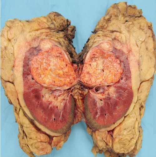
Tumors
Sometimes cysts is able to grow malignant tumors. Unfortunately, this pathology occurs for a long period of time without symptoms only when the tumor, reaching a large size, compresses the vessels within the kidney through the sinus, appears symptoms.
The majority of neoplasms for a long time asymptomatic
However, all the symptoms are typical for diseases of the genitourinary system and highlight some of them characteristic for the tumor is not always possible, only the appearance of increasing intoxication, reducing the amount of urine and extreme pain, unable to force the doctor to suspect the formation of a tumor.
The characteristic feature of the study of the kidneys on the ultrasound, for suspected malignancy, will diffuse heterogeneous changes in the parenchyma of the organ. This feature is explained by the fact that a clear outline tumor formation will not have, growing gradually, it captures new areas of the parenchyma, occasionally moving to adjacent organs and vessels. So when she saw on the monitor screen of the inhomogeneous changes in the body, you should think about Oncology.
Kidney paired organ, therefore, to compare and to exclude diseases in other organ always are screened on both sides. In suspected cancer of the kidney is assigned to a number of other studies (intravenous urography, MRI, CT, radioisotope examination) and only then to put the final diagnosis.
Benign tumors of the sinus of the kidney
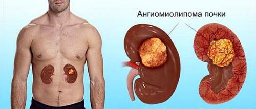
There are also benign tumours of the sinus of the kidney. Types of lipomas that occur in the thickness of adipose tissue that fills the sinus of the kidney varied (angiomyolipoma, lymphangioma,fibroma, etc.)
Lipoma has many varieties
Typically, a lipoma is formed from adipose tissue, which is located in the sinus and serves as a seat and latch for passing vessels and ureters. Has a tendency to rapid growth, but has no ability to invade neighboring tissues and organs.
The size of the lipoma is growing rapidly, constricts blood vessels and the ureter, thereby obstructing outflow of urine. Up to this point the presence of a tumor in the sinus are asymptomatic.
Such tumors as angiomyolipoma, sprouting in the capillaries, gives significant hematuria. Grows to small size. Even the small size of this tumor can cause gross hematuria.
Lymphangioma – numerous tumors, sometimes they can be several different sizes, sprouting in lymphatic vessels, contain large amounts of lymph. They often form in the renal sinus.
Unfortunately, nowadays, diseases of the genitourinary system are not uncommon and not always easy to correct diagnosis, many of them are diagnosed with difficulty. Only a complete, multifaceted examination using modern medical equipment will help to establish the pathology in the body.

At this time I am going to do my breakfast, afterward having my breakfast comingover again to read additional news.
When I initially commented I clicked the “Notify me when new comments are added” checkbox and now each time a comment is added I get several e-mails with the same comment. Is there any way you can remove me from that service? Bless you!
After reading your blog post, I browsed your website a bit and noticed you aren’t ranking nearly as well in Google as you could be. I possess a handful of blogs myself, and I think you should take a look at “seowebsitetrafficnettools”, just google it. You’ll find it’s a very lovely SEO tool that can bring you a lot more visitors and improve your ranking. They have more than 30+ tools only 20$. Very cheap right? Keep up the quality posts
Good piece – we need to read more like this, as most info about this topic is generalized. You provide real insight to people.
You come up with some insightful points within this article, but aren’t you generalising something fundamental?
I truly enjoy looking through on this web site , it holds superb content .
Parasite backlink SEO works well 🙂