Magnetic resonance imaging (MRI) is a method of study of the human body, implying the use of magnetic radio waves and a computer which displays a processed image.
What is MRI and why we need it
The principle of operation of the imager – creating a magnetic field through which hydrogen atoms are converted into radio waves.
This allows the protons of the molecules in the body to create a signal and send it to the receiving device of the tomograph, and then the computer converts the image of the structure of the human body. The images obtained are examined by experts. In some cases, a contrast agent, to improve the detail and accuracy of the image.
Magnetic resonance imaging allows to examine in detail the problem areas of the human body, sacral and other parts of the spine, heart, blood vessels, urogenital system, etc. of Radio waves in an MRI, unlike x-rays, are absolutely safe for health, as the scan does not imply the use of dangerous radiation.
When you need to do an MRI
MRI usually pass on its own initiative or at the direction of a physician to localize and to specify the disease or problem or to Refine the diagnosis.

Using the initial examination with magnetic resonance imaging can directly detect or exclude tumour or the effects of trauma (both new and old). MRI allows to reduce the time spent on different examinations by different doctors to the minimum and immediately get a referral to a specialist or for hospitalization.
To do tomography is possible as many times per year as necessary, despite the fact that radiological methods of the body such as fluoroscopy, computed tomography, x-ray Issledovanie other, preferably, not more than once a year.

Cases in which diagnosis using magnetic resonance imaging:
- problems with the nervous system, tumors;
- morphological changes of the brain and people with Alzheimer’s;
- diseases of the pituitary gland;
- diseases of middle and internal ear;
- heart disease;
- vasoconstriction;
- damage and infection of the joints, bones;
- diseases and tumors of the kidneys, pancreas, lungs, liver, etc.;
- breast cancer;
- problems with the genitourinary system.
MRI can be called the most effective and modern method of research of all departments of the spine, including the sacral region that is most susceptible to disease, so it is worth noting its advantages over other methods:
- images obtained from the scanner have a greater detail;
- received CT information is completely accurate;
- high level information of the images;
- examination of his back is completely harmless to the body;
- the absence of pain in the study;
- a large field of application.
Using magnetic resonance imaging and other sacral spine can diagnose not only bone structure, but also blood vessels, and soft nervous tissue in the spinal cord, as well as:
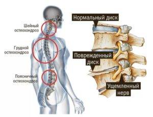
- osteochondrosis of the various divisions of the spine, protrusion and herniated discs;
- injuries and disorders of the statics of the spine (fractures, dislocations, instability vertebrae, scoliosis);
- congenital problems with the development of the spine, displacement of vertebrae (spondylolisthesis);
- hemangioma of the spinal cord;
- infectious diseases of the spine;
- disorders of the lumbosacral spine;
- stenosis, osteoporosis, tumors.
Preparing for MRI of the spine
As the MRI due to the strong influence of the magnetic field, it should observe some simple precautions. Required to remove all clothing containing metal, empty your pockets of all metal objects.
The most important requirement in preparation for MRI sacral and other parts of the spine is the absence of metal implants and prostheses. This requirement must be met as magnetic fields can cause malfunction of the devices in the body, and in the case of dentures could cause serious injury. Also imaging is contraindicated when wearing braces, pacemakers, vascular stents and clips.

Preparing for MRI of the spine is an integral part of the procedure. Only with proper preparation can be guaranteed absolute safety and the safety of magnetic resonance imaging.
The procedure for completing the MRI
MRI is performed by experienced specialists, harmless to humans and does not require any training, if you meet the requirements. The patient should take a horizontal position on the couch, then he fixed my hands and my head to avoid movements that can interfere with obtaining clear images. Couch enters the ring tomograph in such a way that the studied part of the body was in a zone of its action. After that comes the scanning (rotating the device around the scanned area).
How long is an MRI
The time taken for examination, may be very different, it all depends on the scanned area, and from the pictures what quality need to get. For example, a CT scan of the lumbosacral spine could be performed as two minutes, receiving the output images of poor quality, and for half an hour, but in this case, pictures are much more detailed and clear.
Now on MRI is the most important thing: for the diagnosis of sacral spine, and other parts of the body, this method is the most modern, safe and informative.
Magnetic resonance imaging is a complicated and precision instrument. Pictures (tomogram), obtained by MRI, are a comprehensive resource useful for both the patient and physician information. The only disadvantage of this device is that it is not able to find the problem and assess the information received, but it is the work of doctors.


Hi, just required you to know I he added your site to my Google bookmarks due to your layout. But seriously, I believe your internet site has 1 in the freshest theme I??ve came across. It extremely helps make reading your blog significantly easier.
I truly love your website.. Great colors & theme. Did you create this site yourself? Please reply back as I’m trying to create my own blog and want to know where you got this from or just what the theme is called. Thank you.
Wow! This blog looks just like my old one! It’s on a completely
different subject but it has pretty much the same layout and design. Excellent choice of colors!