More than half of the population sooner or later have symptoms of lumbar osteochondrosis. With age, the person wears out the intervertebral cartilaginous tissue, which leads to various diseases of the spine.
Lumbar osteochondrosis, if left untreated and does not take any measures to eliminate the symptomatic pattern, can be a serious problem for the patient.
Such damage to the spine affects the mobility of a person, and the peculiarity of the course of the disease prevents a full life.
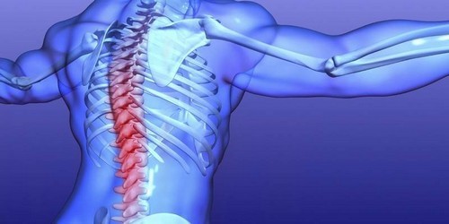
Pain in the spine
Depending on the degree of development of the pathological process in the vertebral column, the person will have different symptoms. As with any other disease, the more developed the destructive process, the brighter the clinical picture. Osteochondrosis in the lumbar spine is formed due to a strong and prolonged load on the spinal column. Accordingly, the most obvious symptom in this case – the inability to perform the usual physical activity. In addition, there is a difference in the clinical picture with each degree of lesions of the lumbar spine.
The main symptoms of the lumbar osteochondrosis form a single picture of the disease, which can almost immediately be recognized, without diagnostic measures. The patient’s complaints to the doctor and the visible symptoms in the acute period speak for themselves:
With the development of pathological destruction of the vertebrae in the lumbar region with osteochondrosis, the patients develop the strongest pain syndrome. The more destruction occurs, the less a person tries to make any movements, which leads to secondary signs of osteochondrosis. Patients describe the pain as acute, and often in complaints, doctors hear such an expression as “a count in the lower back.” It is this symptom that catches the patient at home, when the vertebrae in the lumbar region are displaced, pinching the roots of the nerve endings.
Forced position of the body in pain a person takes involuntarily. This is the instinct of self-preservation, the patient tries not to change the posture in which he was overtaken by the pain syndrome, as when trying to straighten, sit or lie down, it is greatly strengthened. Often people note the pain radiating to one or both legs, to the groin or lower abdomen.
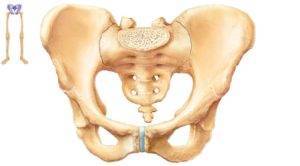
Low back osteochondrosis
Numbness and chilliness in the lower limbs also occur with damage to the nerves, and, as a rule, this symptom increases with the progression of the disease. Numbness comes in the position lying on the back and if a person sits in one pose for a long time. Excessive physical exertion, overstrain of the back muscles, trauma can lead the disease into an acute form, in which all the symptoms will intensify and the destruction of the cartilaginous tissue of the vertebrae will be even more intense.
The onset of pain syndrome is the main symptom of low back osteochondrosis. This is the main complaint of all patients, and it is this problem that is fundamental in the initial stage of diagnosis. Localization and irradiation of pain in either side of the lower part of the body shows how much the cartilaginous tissue is destroyed, as well as the area of nerve root seizure. Deterioration of the process is manifested by a significant increase in the symptomatic pattern.
Secondary signs
When a patient develops an intense pain syndrome, a person always tries to avoid pain intensification by any means, often taking a forced position of the body. Symptoms of the lumbar osteochondrosis lead to the fact that the patient makes fewer movements, avoids physical exertion.

Secondary signs of osteochondrosis begin to appear at a time when a person due to illness manifests physiological disorders. For example, numbness of the legs due to impaired movement of impulses along the nerve arches, as well as changes in sensitivity, indicate degenerative changes in the lumbar vertebrae.
An effective remedy for pain and for the treatment of joints has been found:
- natural composition,
- without side effects,
- effectiveness, proven by experts,
- quick result.
The appearance of visible symptoms of the disease, such as a hump, curvature of the spine and an increase in osteophytes, shows how much the structure of the vertebrae and intervertebral discs is changing. Their compaction and thickening leads to significant changes in motor functions.
Methods of diagnosis
The beginning of diagnostic measures begins with the examination of patients by such specialists as the neurologist and orthopedist. Already at the initial stage it is possible to diagnose the patient on the basis of data of anamnesis and primary examination, since the osteochondrosis of the lumbar region has clear symptoms that are visible without special equipment.
The main methods of diagnosis of lumbar osteochondrosis:
- radiography;
- CT scan;
- Magnetic resonance imaging.
For more accurate diagnosis, laboratory and instrumental methods of investigation are used. Moreover, laboratory methods are not aimed at diagnosing osteochondrosis, but are used to identify possible inflammatory diseases of the body, which can significantly complicate the picture of the disease and further treatment, which is often observed in patients.
Important! During the exacerbation of the disease, the diagnosis of lumbar osteochondrosis is not performed, since a person is not able to take the necessary position of the body to perform the pictures because of the intense manifestation of the pain syndrome. First, you need to reduce the pain so that the patient can move freely.

Instrumental diagnostic methods consist in the application of various procedures by which it is possible to see the state of the bones of the spine and cartilaginous tissue. X-rays are expelled in several projections to assess the condition of the vertebrae, the presence of deformity, the shape and height of the intervertebral space, the growth of osteophytes.
In order to assess the condition of the intervertebral openings and joints, the x-ray is performed in oblique projections. If the patient has impaired mobility of the spine or pathological mobility (one that should not normally be), the pictures are produced in the bent and straight position of the spine.
Currently, the x-ray method for the study of osteochondrosis is used much less often, because it is less informative than other methods of research.
Scanning the spine with the help of tomographs allows you to see the damage picture in more detail. Computed tomography is the most preferred diagnostic method in comparison with x-rays. CT uses the same X-ray radiation, but it is much smaller, which makes the study much safer for the patient, and it can be produced more often. The received pictures are more informative: with their help it is possible to determine which structures damaged the disease.

Magnetic resonance imaging is performed with the help of electromagnetic radiation, which is absolutely safe for a sick person. This method of diagnosis is used to display the most accurate picture of spine injuries. MRI allows you to consider any, even the smallest changes in bone or cartilaginous structures, to detect damage, to assess the condition of tissues.
Thanks to magnetic resonance imaging it is possible to observe changes in the damage pattern in the dynamics of the treatment, since this method does not produce irradiation per person and can be used more often than other methods of investigation. When planning surgical interventions, the MRI allows the traumatologist to develop an accurate operation plan and prescribe treatment.