
Chondrophora of the larynx is inflammation of the cartilage of the larynx and pericentre – thin connective tissue film covering the laryngeal cartilages.
Pathology is in the form of banal catarrhal lesions, and purulent.
The disease can be detected very unpleasant, as when it pronounced symptoms significantly suffers from the act of breathing.
General data
Chondrophora of the larynx is less common than other diseases and pathological States. The overall incidence is about 1-2 cases per 10 000 population. But the development of severe septic complications and their critical consequences encourages clinicians to exercise caution in relation to this disease.
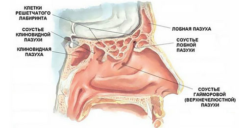
In most clinical cases, there is inflammation of the arytenoid cartilage and the cricoid, rarely the thyroid, rarely affects the epiglottis. The last fact is favourable to the human pathology of the epiglottis, because of its location can lead to quickly advancing to asphyxia (suffocation).

Most often, the described disease is diagnosed in the age group of 30 to 50 years. Children of any age can get sick pretty rarely. Pathology affects with approximately equal frequency in patients of both sexes.
More than 35% of all clinical cases of chondrodermatitis larynx appear complications such as stenosis (narrowing) of the larynx. This means that the relief of acute symptoms relieves the patient from unpleasant effects of this disease.
Reasons
As the throat flow of infected air, and its structures are not sterile, chondrophora of the larynx has a typical infectious-inflammatory in nature. Isolated pathological process by only pericentre or just the cartilage of the larynx does not occur.

The immediate cause of the described disease are infectious agents. As in the majority of cases, infection of the respiratory tract, the larynx handoperated often causes nonspecific pathogenic microflora is a “universal” pathogen of infectious-inflammatory process which may develop in various organs and tissues. Most often these are infectious agents, such as:
and some others.
Less chondrophora of the larynx may occur if the penetration of tissue specific agents of infection – it’s basically:
and some others.
Described the disease rarely can cause:
Chondrophora of the larynx are a source of infectious agents, living in chronic foci of infection in the body until some time residing in a state of inactivity or of low activity.
It is not uncommon for the occurrence of laryngeal chondrodermatitis only one infectious agent is not enough – pathogenic infection in the cartilaginous tissues of the larynx often occurs only on the background of some of the factors contributing to the development of this disease.
Most often it is:
- traumatic injuries of the larynx;
- concomitant diseases of an infectious nature;
- impaired immunity;
- the weakening of the body as a result of previously deferred or diagnosed at the moment of serious diseases, conditions or operations;
- ecological trouble of environment;
- radioactive impact.
Traumatic injuries of the larynx that contribute to the development of chondrodermatitis, this injury:
- medical origin;
- non-medical origin.
The first group of factors include the manipulation of:
- diagnostic;
- treatment.
Traumatic with subsequent appearance of chondrodermatitis can detect such diagnostic procedures as:
- laryngoscopy – examination of the larynx using the laryngoscope (one of the types of endoscopic equipment);
- a biopsy of the larynx – fence part of it is tissue for subsequent laboratory diagnostic;
- bronchoscopy – inspection of the bronchial tree with the help of a bronchoscope (a kind of endoscopic techniques), which is introduced into the bronchi through the larynx.
Trauma of the larynx and the subsequent development of its chondrodermatitis possible when carrying out such medical procedures as:
- incorrect probing of the esophagus – introduction lumen extends special metal rods when because of a violation of the manipulation techniques the bougie may enter the lumen of the larynx and injure its wall.
- careless removal of a foreign object in the larynx, trachea, bronchi and esophagus;
- conicotomy – the creation of an artificial opening into the laryngeal wall resulting in asphyxia (suffocation). This is one of the most frequent causes of trauma to the larynx, as is done in urgency, when time pressure increases the risk of erroneous actions;
- any surgical intervention on pharynx, trachea, esophagus.
Trauma of the larynx that is not related to medical procedures is observed in such circumstances as:
- injury during class power sports – Boxing, wrestling, hockey, football and other;
- road traffic accidents;
- natural and man-made disasters;
- intentional strikes with his fist or improvised subjects in the neck;
- damage to the cartilage of the larynx at the attempt of suffocation or hanging (including suicide);
- the stabbing in the neck cutting or piercing objects;
- bullet and shrapnel wounds.
Well as injuries of the larynx, contributing to the development of chondrodermatitis include burns:
- the chemical is in contact with the inner surface of the wall of the larynx corrosive chemicals;
- thermal – as a result of exposure to open flame, hot liquids or steam.
Diseases of infectious nature which contribute to the development of chondrodermatitis can develop from LOR-structures from other organs and tissues.
Most often it is diseases such as:
- laryngeal sore throat – inflammation of the islets of lymphoid tissue scattered throughout the inner surface of the larynx;
- influenza – a contagious infection triggered by different types and strains of influenza virus;
- submucosal abscess – limited abscess that develops under the mucous layer covering the inner surface of the walls of the larynx;
- laryngitis – inflammation of the tissues of the pharynx;
- pneumonia – inflammation of the parenchyma of the lungs;
- erysipelas – an infectious lesion of the superficial layers of the skin;
- typhoid fever – an acute intestinal infection caused by Salmonella;
- typhus – an acute infectious disease caused by Rickettsia and is characterized by a specific rash;
- tuberculosis;
- syphilis – a sexually transmitted disease.
Impaired immunity, which increases the risk of chondrodermatitis of the larynx can manifest in the form of a temporary weakening of the immune system, so the form is constantly observed immunodeficiencies – congenital or acquired.
Chondrophora of the larynx are often diagnosed in immunocompromised patients in the following cases:
- after suffering a major surgery (especially abdominal – the abdominal and thoracic cavity);
- after a long stay in a serious condition in the intensive care unit or intensive care;
- amid protracted chronic diseases – hepatitis, pancreatitis, systemic connective tissue diseases, rheumatic diseases and many others.
The development of chondrodermatitis of the larynx contributes to permanent (permanent) stay in the conditions of polluted atmosphere, in particular when living in an industrial region.
In some cases the contributing factor may be the radiation exposure – in particular, due to the radiotherapy for this or any other malignant guidance in the body. Radiation therapy reduces local resistance (resistance) of the tissues including cartilage and perichondrium of the larynx.
The development of the disease
An infectious agent can penetrate into the cartilage and perichondrium of the larynx in different ways, primarily:
- hematogenous (through the blood);
- contact.
First develops an inflammatory lesion of pericentre – namely, from the inner part, which is adjacent to the cartilage. The outer layers of perichondrium are more resistant, therefore, at the initial stage of pathology they observed unexpressed inflammation – it occurs:
- slight redness of the tissues;
- their moderate infiltration (seal);
- insignificant growth of connective tissue.
The inner layers of perichondrium (they are responsible for the growth and vascularity of the cartilage) are more sensitive to inflammatory lesions. Because of this, very soon between the perichondrium and cartilage accumulate exudate – the fluid that came out of the cells and from the lumen of the small vessels, because the permeability of their walls increases inflammation. All these processes affect trophic (feeding) cartilage and their immunological resistance, which has already been reduced on the background infectious process. Therefore, joins the inflammation of the cartilage, which is accompanied by necrosis (necrosis) of the cells and sequestration (literally loss of dead cells of the cartilage).
There are two forms of chondrodermatitis of the larynx:
- primary;
- secondary.
Primary handoperated occurs as an independent disease, which is a direct infection of the cartilaginous skeleton of the larynx. In particular, it is a pathological process that develops as a result of injury and spread of the pathogen from distant infectious foci.
Secondary handoperated is a complication of infectious processes of adjacent organs and tissues. Mostly they are inflammatory processes in the mucous membranes of the tonsils, trachea, bronchi, and so on.
There is a separate form of chondrodermatitis depending on the clinical course:
- purulent (aka abadimulia);
- sclerosing (aka fibrous).
In the first case, the inflammatory lesions of the cartilage and begins as a catarrhal pericentre (simplified inflammatory process), further in the progression it becomes purulent, forming an abscess – a limited abscess.
In the second case, the inflammatory process is the cause of development of connective tissue. In the cartilage and the perichondrium are observed sclerotic changes, perichondrium when it grows, the end result scars are formed from connective tissue.
Depending on where the pathological process, handoperated happen:
- domestic – while in the pathological process mainly retracts the inner surface of the cartilage and pericentre from the lumen of the larynx;
- external inflammatory process develops in the outer perichondrium, which leads to more pronounced breaches in the front of the neck.
The symptoms of laryngeal chondrodermatitis
During the initial chondroitinase the clinical picture develops acutely. The symptoms are the following:
- fever – simultaneously observed hyperthermia (increased body temperature) and chills. Hyperthermia may reach 39.5 C to 40.0 degrees Celsius;
- very marked weakness;
- bottled intense headaches;
- breathlessness inspiratory nature, sick hard to breathe, exhale may remain unchanged.

When the secondary chondroitinase larynx pathological process develops gradually. The symptoms are the same as that of the primary form, but not as severe, more moderate, can sometimes be absent.
The clinical picture of the outer chondrodermatitis the following:
- pain syndrome;
- after some time – the formation of an abscess in the form of small dense formations, painful at palpation.
Characteristics of pain:
- localization – most often on the anterior surface of the middle or upper third of the neck;
- distribution across the surface of the neck, in the parotid region;
- by nature blunt, the formation of an ulcer – twitch;
- intensity – moderate, formation of an abscess – a fairly noticeable;
- the appearance – occur with coughing, swallowing and talking, growing as the transformation of infiltrate in the abscess, worse when bending and turning the head.
Characteristics of education:
- shape – round;
- size small, from 1 cm in diameter;
- consistency – dense, which after a time softens;
- sensitivity, painful at palpation.
As the transition process in purulent skin in the neck becomes thinner, becomes bluish, then yellow-brown. Purulent contents can break through, forming a festering fistula (abnormal passage). As soon as the abscess breaks through the General condition of the patient immediately improves.
Internal handoperated more severe because developing stenosis of the larynx. Clinical symptoms are the following:
- inspiratory dyspnea;
- tachypnea;
- stridor (wheezing);
- hoarseness. Is a typical symptom of the described pathology;
- changing the timbre of the voice. It can be so pronounced that the voice becomes unrecognizable;
- respiratory failure occurs when the progression of the pathology;
- bruising exposed parts of the body – the tips of the nose and ears, fingers;
- fatigue, even if the loads remain the same.
Sometimes when perichondritis laryngeal stenosis develops so intensively that there is a need for urgent tracheostomy – forming an artificial opening in the trachea below the narrowing.
If the internal chondroitinase breaks through the abscess, you experience the following symptoms:
- a sudden bout of pronounced coughing;
- expectoration of a large quantity of pus.
After breaking internal abscess the General condition of the patient is significantly improved, all of the above described symptoms could be subtle or disappear.
Diagnosis
Most often, the diagnosis is based on complaints of the patient and inspection data. Additional methods of examination carried out to clarify the changes and detection of complications.
Results of physical examination:
- during the examination – in protracted course of the disease is marked pallor of the skin, visible mucous membranes and fingertips with cyanotic tinge, the patient is panting, it is noted the presence of significant respiratory noise;
- auscultation (listening with a stethoscope) to hear noisy breathing.
Apply following instrumental methods:
- indirect laryngoscopy – examination of the larynx using the laryngoscope (a variety of endoscope). Marked narrowing of the lumen of the larynx, redness and swelling of the mucous membranes;
- radiography of the neck – used to detect changes in outdoor chondroitinase. In the presence of abscess on x-ray picture shows the deformation of the cartilage of the larynx, as well as a shadow (it forms an abscess), often with a horizontal fluid level (due to the pus);
- computed tomography of the neck (CT) – computer sections provide more information about tissue at the site of lesion, to identify the ulcer, to study its characteristics;
- magnetic resonance imaging of the neck (MRI) – challenges and opportunities the same as CT.

Laboratory methods, which are used in the diagnosis of chondrodermatitis is:
- General analysis of blood – there has been a sharp increase in the number of leukocytes and ESR;
- Wasserman – it is positive when chondrodermatitis syphilitic origin;
- the Mantoux test is performed to confirm the tuberculous origin of chondrodermatitis;
- bacterioscopic study under a microscope being studied purulent contents obtained by expectoration, it identifitseerida the pathogens that caused the development of chondrodermatitis;
- bacteriological examination – make odalengo content seeding on nutrient media, growing colonies determine the causative agent and its sensitivity to antibiotics.
Differential diagnosis of
Differential diagnosis of chondrodermatitis larynx often carried out with such diseases as:
- laryngitis – inflammation of the mucous membrane of the larynx;
- esophagitis – inflammation of the lining of the esophagus;
- abscess of the soft tissues of the neck.
Complications
Most often, when chondroitinase of the larynx develop the following complications:
- stenosis of the larynx – its contraction;
- chronic hypoxia – lack of oxygen in the blood due to disorders of the airway;
- aspiration pneumonia – inflammation of lung parenchyma due to the ingress of purulent content in the breakout abscess;
- asphyxia – a state of suffocation when injected pus into the respiratory tract;
- cellulitis of the neck – diffuse purulent lesions of the soft tissues;
- intracranial infectious complications;
- mediastinitis – inflammation of the complex of organs and tissues, which are located between the lungs;
- sepsis – spread of infection throughout the body with the formation of secondary infectious foci.

Treatment of laryngeal chondrodermatitis
Therapeutic activities are carried out exclusively in the hospital environment. Treatment depends on chondrodermatitis of the larynx, its severity, presence of complications.
Purpose in pathology without the formation of abscess the following:
- massive antibacterial therapy. Is assigned based on the sensitivity of pathogens. To determine the sensitivity prescribe broad-spectrum antibiotics;
- anti-inflammatory drugs;
- antihistamines;
- infusion therapy for the elimination of intoxication syndrome. Intravenously administered saline and protein solutions, electrolytes, glucose, blood serum;
- vitamin complexes;
- physical therapy is assigned in the absence of an ulcer. Proven UV, UHF, microwave, phonoelectrophoresis, monohalobenzene with potassium iodide or calcium chloride.

If an abscess has formed, surgical intervention is required in his opening, emptying and drainage. Often, before the operation is performed a tracheostomy – in this case, can be carried out without anaesthesia threat of penetration of pus into the lungs.
Prevention
The risk of chondrodermatitis of the larynx can be reduced if you adhere to these recommendations:
- avoidance of situations that constitute a trauma of the larynx;
- treatment of opportunistic infections;
- strengthening the immune system and General body strength after diseases and operations.
- avoid staying in polluted environment;
- avoidance of radioactive exposure.
Forecast
The prognosis for chondroitinase of the larynx is favorable, if the disease is identified in a timely manner, assigned adequate treatment.
Prognosis worsens in severe cases, since irreversible changes in the tissues of the larynx, which provoke the failure of respiratory function. In this case, to help the patient is only possible by surgical plastics of the larynx.


The latest Free stuff, freebies, free samples, free trials, prize draws, competitions, discount codes, vouchers, coupons and hot deals from the UK.
Thanks for this helpful information.