Perforation of the tooth is a serious dental problem complicating the treatment process nearly 10% of cases. Why it occurs, how dangerous and how is it treated, can be found below.
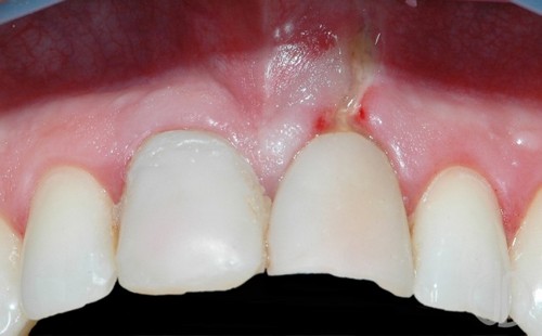
Perforation of the tooth is usually represented by a hole in the tooth structures that can occur due to complications of a mechanical injury or for other reasons. The defect can occur in areas such as the bottom of the dental cavity, the wall element of the jaw or roots.
Perforation tooth hole in tooth structures
According to experts, there are the following causes of disease:
- Individual anatomical features of the canal system of the root. If the channels have a curved structure, to work with them, the doctor will be more difficult. Perforation can happen at the time of the expansion of one of them or when setting pin;
- The carious process destroys tooth unit and it thins the hard tissue, causing the formation of holes;
- Mechanical trauma to the hard tissues (shock, spalling). In this case, the defect can be represented by a crack. Sometimes problems occur when careless actions of a physician (improper distribution of force in the application of instrumentation, which causes excessive pressure on the dental tissues).
There are several factors causing the appearance of the defect:
- The deviation of the Central axis of the tooth in the direction;
- Implementation of medical action through the crown;
- Erasing tissues of the tooth, leading to increased thinness of its walls.
If you have been diagnosed, but the doctor was not timely, we can expect the following consequences:
- Change the anatomical features of the jaw elements;
- The bone loss.
Specialists distinguish the following types of defect depending on its localization:
- Perforation of the bottom cavity. Occurs because of displacement of tooth of the axis (toward the cheek, tongue). Sometimes the problem can be triggered by erasing the chewing surface, during medical events – excessive expansion of the cavity;
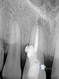
- Perforation of the tooth root. Common type of that require immediate treatment, otherwise, increases the likelihood of destruction of connective tissue (the periodontal). The sooner therapy is begun, the more likely its effectiveness. Sometimes the pathology is associated with the curvature of the roots. To clarify its localization requires x-ray;
- Perforation of the walls of the unit. Mostly caused by sloppy work of the doctor, aggressive action on the element of the jaw during treatment. A symptom of the problem is the abnormal position of the instrumentation, the bleeding, the patient may feel a sharp pain.
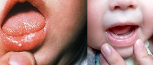
Symptoms
Almost immediately after the problems the patient feels a sharp pain, maybe bleeding from wounds. On examination, the physician notes abnormal occurrence tool (it “falls”), the presence of vaginal bleeding, sees the wound area.
If the pathology is chronic, it may not be evident, but its presence is evidenced by periodic bouts of aching pain.
Modern methods can effectively treat dental hole walls. However, if the affected cavity, it is important to consider the size of the damage. Filling will yield positive results only with a small hole (2 mm), in other cases likely to be filling materials in the connective tissue and infection.
Therapy of this pathology may be of therapeutic or surgical in nature.
Treatment
It can be used to eliminate the holes to 3 mm. Perhaps the elimination of perforation of the tooth root if it is localized in the middle or the coronal portion. Disadvantage – the possibility that the filling material in the periodontium with subsequent inflammation.
To cure the defect in the presence of a cemented pins and kultovyh tabs and to eliminate the granulation, rebuild tissue, bones treatment of perforation of the tooth will not help, the necessary surgical procedures.
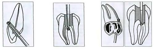
Surgical treatment
The main goal of surgical treatment of perforation of the tooth in this case is its reliable sealing using glass ionomer cement (elements covering the bone are filled with osteoplastic material). The doctor uses a biocompatible material that should be resistant to damage and allows you to tightly attach it to the field of sealing.
Composites, unlike glass-ionomer cements do not meet the requirements necessary for the elimination of perforations (compatibility with the tissues of the patient, high strength and perfect adhesion, sufficient adhesion), therefore do not apply.
Prevention
- The use of modern safe equipment during inspection and treatment, accurate for dental work;
- Creation of conditions for the review of cavity of the jaw units when the Dr;
- Constant control of anatomical structure, location, and unit condition. Is provided by radiography after each complex manipulations;
- Mandatory removal of artificial crowns to the start of endodontics.
Possible complications
- Granuloma. Inflammation can lead to vesicle with purulent contents. Further pathological focus moves to the nearby area and even to the periosteum, there may be fistulous passages;
- Cyst. Appears as a consequence of lack of care granuloma. The inflammatory process can develop to go beyond the mouth, affecting other organs. Increased risk of loss of the unit, the process can stop only qualified medical assistance. After removal it is necessary to exclude the presence of pathological elements, since otherwise the recurrence of tumors;
- Broke off the root. Can happen if the hole seriously damages the structure of the unit. Leads to inflammation, complication of chewing and speech function can cause loss of units or jaw misalignment. Broke off, confirmed by radiography;
- The root in the gum. If the removal was done poorly, the remaining fragments will lead to infection and swelling, severe pain.