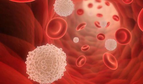
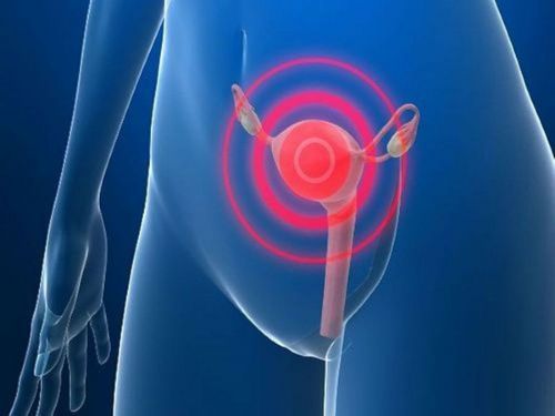
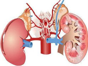
Erythrocyturia – literally means “red blood cells in the urine”. The same term “hematuria” (blood in the urine), which is more common in the medical environment.
A symptom is considered to be very important in practical activity of doctors of different specialties. The reasons may not necessarily escape in disease of the urinary organs.
Modern diagnostics tries to identify the blood cells in the urine, but also to establish where they came from. This indicates the defeat of the body. Therefore, we conducted a study in both the number and qualitative properties of red blood cells in their biochemical composition.
What is considered eritrotsituriey?
It is estimated that during the day, a healthy person passes into the urine, about 2 million red blood cells. In the clinic, the determination is carried out according to the number in the field of view of the microscope.
Evaluation standards by different specialists is a little different, but the majority accepted as the norm to be considered;
- women – up to three red blood cells;
- men 0-1.
In children the cellular level depends on the age. The body of a newborn human is doing a tremendous job for the transition of hematopoiesis from the parent on their own. Those red blood cells that remain from the time of intrauterine life, are destroyed.
This process is usually accompanied by physiological jaundice and uric acid diathesis. In the urine detected up to 7 “old” erythrocytes in sight. How long lasts the adaptation of the child depends on his individual characteristics.
Note that full activation of renal filtration function occurs in 2-3 years. So the baby is 12 months of age the norm is up to 5 cells per field of view.
After three years, a healthy child establishes common indicators:
- for girls – 0-3;
- boys – one in sight.
In the analysis it is possible to estimate such quantities as “single”.
The eritrotsituriey is any condition in which the urine is detected the number of cells more than the norm.
Micro and gross hematuria, the differences
Quantitative feature of erythrocyturia is characterized as:
- microhematuria – the patient does not notice the changes in urine color, it remains a straw yellow, but when microscopy laboratory sees a number of red blood cells in field of vision, exceeding established norms, usually detected during preventive examination or turning in the opinion of the patients not associated with kidney disease;
- gross hematuria – erythrocytes cover all visual field, there are 100 or more, the color of urine becomes reddish-brown, patients themselves see unusual changes sometimes appear on the toilet blood clots.
It is impossible to judge about the intensity of erythrocyturia only in color of urine
Reddish shades arise:
- by eating beets;
- receiving significant doses of medications containing Aspirin and Analgin;
- during the course of treatment with Cyanocobalamin (vitamin b12).
As the red blood cells get into urine?
The presence of erythrocytes in the urine is always alarming the doctor and suggests pathological changes. This is due to the fact that they are constantly in the bloodstream. Urine for unusual cells habitat.
The most numerous blood cells grow in the bone marrow, the spine, the bones of the skull, ribs. Babies grudnichkovye age, there is an additional source – the bones of the limbs. Almost 90% of the mass of cells is hemoglobin is a complex biochemical compound is a protein with iron. In connection with large size, red blood cells cannot pass through the membrane of the glomeruli, if it’s not destroyed.
To protect from mixing of blood with the urine in the body created by physiological barriers.
These include:
- quite dense and elastic vascular wall;
- a strong fibrous capsule of the kidney;
- the basement membrane of the glomeruli with selective permeability;
- epithelial and muscle layers in the lower urinary tracts (bladder, ureters, urethra).

Physiological or temporary (transient) hematuria possible at elevated pressure leading to the capillaries of the glomeruli, it pushes the red blood cells through the basal membrane
Pathological mechanisms of erythrocyturia can be the following violations:
- increased permeability of the blood vessels that supply blood to the organs of the urinary tract, it is possible as a result of local inflammatory reactions, traumatic injury, tumor lysis;
- the destruction of basal membrane of renal glomeruli in the nephritis of various etiologies of toxic exposure to toxic substances, certain drugs;
- with the General stagnation in the veins of the pelvic organs (phlebitis, growing uterus in pregnancy, mechanical compression by tumor, or hydronephrosis of the kidney).
Causes of erythrocyturia can be viewed from the point of view of violation of mechanisms for the protection and development of the specific pathology. Classification of pathological causes is subdivided according to the type and level of injury in extrarenal and renal.
When there is a physiological reason?
Cause of red blood cells from the blood vessels in the urine can be a reaction to physiological processes. While erythrocyturia characterize as microhematuria. Never macroparasite with visible bleeding. Physiological microhematuria always disappears on its own after the expiration of its causing factors.
A moderate increase of erythrocytes in the urine:
- in connection with prolonged exposure to UV rays, overheating in the sun, after the “sun stroke”;
- on the background of alcohol abuse, even a single;
- with overeating meat heavy, fried, spicy and salty foods;
- after steaming in the bath;
- in athletes under intense training, the soldiers “March” hematuria;
- after lifting, heavy work;
- people often suffer from stress reactions and stressful life situations.
Most sensitive to these factors, children. The combination leads to even more complicated conditions and disrupts the mechanisms of adaptation. Surveillance for several years, men with previously identified eritrotsituriey after a long forced March showed the absence of disease of the kidneys.

People with physiological hematuria is necessary to monitor the urine, to conduct a full examination. It is known that in pathological mobility of the kidney is also possible temporary hematuria. It is associated with the vertical position of the body, disappears in the supine position. Physiological causes should be differentiated disorders that occur when collecting urine for analysis. If the scheduled urine test, you should exclude all the above factors of influence for 2-3 days before collection.
Women should not have to provide a urine sample within a week after menstruation, because it is possible to hit in a container of blood from the vagina
The necessary hygienic cleaning eliminates the effect of the bleeding of hemorrhoids, fissures of the rectum. Failure to comply leads to false data and creates a reason for a second study.
Extrarenal erythrocyturia (hematuria)
In a group of extrarenal disorders included any pathology with hematuria not related to direct injury of tissue of the kidneys. It can be as diseases of the urinary organs, and diseases of the heart, blood, vessels.
Of diseases of urinary organs, hematuria is often accompanied by:
- bladder tumors – cancer of this localization is most common in older men;
- adenocarcinoma of the prostate in young and middle-aged men;
- traumatic injury of the ureters, bladder, urethra in fractures of the pelvis injuries;
- urolithiasis – discharge of stones cause damage to the blood vessels of the mucous membrane of the ureters;
- tuberculosis of the bladder.

Hematuria is caused by lesions of other organs and blood vessels. This mechanism is accompanied by:
- disease blood clotting disorders (hemophilia, thrombocytopenia);
- vasculitis and diathesis (hemorrhagic kapilliarotoxicos);
- decompensation cardiac defects, diseases of the myocardium, contributing to the stagnation in the veins of small pelvis;
- prostate disease in men, cervical erosion, uterine bleeding in fibroids in women;
- infectious fever with high body temperature;
- hypertension of any origin;
- negative properties of drugs in the treatment of sulfonamides, anticoagulants, high doses of vitamin C, Hexamine, overdose, individual sensitivity, the wrong combination in the combinations, reinforcing.
Chronic hemorrhagic cystitis with ulceration of the bladder wall often affects women, inflammation increases the permeability of the vascular wall of arteries and veins
Erythrocyturia renal (hematuria)
Mechanisms of renal hematuria associated with exposure to structures of the body.
These include:
- the destruction of the basement membrane of the glomerular membrane is affected in infectious, autoimmune nephritis, glomerulonephritis, as a consequence of inflammation of the parenchyma with interstitial nephritis, as a result of compression in amyloidosis of the kidney, end-stage pyelonephritis;
- local disruption of the membrane cause hemorrhagic fever, diathesis, blood diseases;
- toxic effect on tubules and interstitial tissue of poisons in cases of poisoning, medications (Heparin, Cyclophosphamide, Warfarin);
- mechanical squeezing at polycystic kidneys, hydronephrosis;
- the total increased intravascular coagulation on the background of antiphospholipid and disseminated intravascular coagulation syndromes;
- necrosis of tissue parenchyma with hematuria develops when sickle-cell anemia, renal vein thrombosis, kidney infarction, malignant hypertension.
The mechanism of degradation of the glomerular apparatus is the basis of inherited diseases with mutations of the chromosome apparatus.
These include rarely diagnosed pathology:
- Fabry disease;
- goodpasture’s syndrome;
- essential and HCV-associated mixed cryoglobulinemia;
- IgA-nephropathy.
Practitioners in work, adhere to the more simple classification of the causes of hematuria, according to which they are divided:
- in prerenal – having no connection with any renal pathology (infectious disease, poisoning, sepsis, blood disease, heart injuries);
- renal (kidney) is always associated with primary renal disease (all nephritis, a kidney injury);
- postrenal – determined by destructive processes in the ways of the urinary tract (urolithiasis, bladder cancer, cystitis, anomalies of the ureters).
The types of erythrocyturia the clinical course
When monitoring patients, there are the following types of hematuria.
For the duration of the course:
- persistent (permanent) – if you save a few analyses carried out at time intervals;
- recurrent blood cells in the urine it is absent, then occur again.

Depending on the combination with other changes in urine:
- isolated – if other components precipitate in the normal range;
- combination with proteinuria – if analysis, in addition to erythrocytes, elevated protein.
In parallel with the red blood cells in the urine can be detected in proteins
The clinical course distinguish between gross hematuria:
- asymptomatic (silent) – appears suddenly by blood clots on the toilet, excitement patients is justified, because of the need urgently to be examined to exclude tumors of the kidney or bladder;
- with the formation of large shapeless blobs on the background of urinary retention is possible when the source of bleeding is in the bladder;
- with clots worm-like shape indicates the formation in the ureters of casts, the source of bleeding are often the kidney or the pelvis for stones in the kidney, polycystic disease, occurs after an attack of renal colic.
Quantitative and qualitative assessment
In the diagnosis it is important to fix a minimum increase in red blood cells even with microscopic hematuria. For this purpose, methods Nechiporenko and Hamburge. They studied the number of erythrocytes per ml of urine, it also checks the content of leukocytes and cylinders. For evaluation, use the index rules to 1000 cells/ml.
Qualitative characteristics of the composition of red blood cells in urine allows to establish the alleged injury. On this basis, cells are divided into fresh (unaltered) and altered (leached).
The microscopic picture of normal red blood cells characterized by rounded contours with pulled inward at the center. The size of the cells is less than the large leucocytes, but more platelets.

Type and form are entirely dependent on the degree of saturation of hemoglobin.
- Unmodified red blood cells – combined with leukocyturia, are formed in the urinary tract, indicate the presence of stones, polyponic formations, tumor disintegration, tissue necrosis with infarction of the kidney, with possible benign prostatic hyperplasia.
- Leached appearance – formed by the loss of hemoglobin differ wrinkled or annular shape, combined with proteinuria in nephrotic syndrome, acute and chronic glomerulonephritis, kidney damage by toxins or poisonous substances.
Leaching of red blood cells is not always associated with pathology. He goes, if the food contains no alkali components. These foods include buckwheat, nuts, vegetables. In conditions when a person goes to a salt-free diet, noticeably disappears the alkaline reserve. Therefore, the body goes into a mode of “self-reliance” and brings lye’s own cells.
Of the patient is necessary to familiarize with preparation for delivery of the analysis and obtain reliable data
Is it possible to identify the level of lesion urinary organs?
To install the urine the diagnosis is very difficult, one feature erythrocyturia can be a component of different combination of the lesion. Practical experience of the old doctors are still likely determines the center of trehstakannoy the sample.
The patient needs to urinate in the morning consistently without a break jet in 3 capacity. Each portion is tested on erythrocyturia.
The conclusion is based on the following options:
- if the red blood cells detected only in the first portion (initial hematuria), the pathology must be sought in the urethra (inflammation stuck microlite, tumor);
- erythrocytes increased in the third portions (terminal hematuria) means the probability of diseases of the bladder inflammatory or neoplastic nature of crushing stone in the neck;
- erythrocytes completely in all tanks (total hematuria) – it makes sense to look for abnormalities of the kidneys or ureters.
How is diagnosis of erythrocyturia?
Doctors pay attention to the combination of gross hematuria with dysuria.
Such features are characteristic:
- for haemorrhagic cystitis;
- urolithiasis;
- tuberculosis of the kidneys and bladder;
- parasitic inflammation.
Combination with lots of protein, cylindruria serves as an indicator of nephrotic syndrome with glomerulonephritis, interstitial lesions, amyloidosis, chronic kidney failure.
Hematuria laboratory study with:
- special test strips for hemoglobin;
- microscopy of the urinary sediment;
- according to the method Nechiporenko (1 ml), Amburgo (1 minute).
For the differential diagnosis of erythrocyturia conduct:
- urine analysis (important can be protein, white blood cells, cylinders, bacteria, salt crystals);
- biochemical tests of blood (as an indicator of nitrogen metabolism creatinine, electrolyte composition, alkaline phosphatase);
- immunological blood samples (indicate the role of autoimmune processes by increasing IgA, cryoglobulins, antinuclear antibodies).
The use of phase-contrast microscopy identifies distinctive features of glomerular (glomerular) hematuria from aglomerarea:
- glomerular – is characterized by differences in size and form more than 80% of the erythrocytic cells (dimorphism), they have adorandote shells and uneven contours;
- aglomerarea – more than 80% of the cells size and shape the same (isomorphism);
- combined form.
To confirm the diagnosis, the patient is administered:
- zitostaticescoe study (allows you to identify the one or two – sided hematuria);
- intravenous excretory urography;
- ultrasound examination of the kidneys;
- computed tomography.
Rarely used retrograde pyelography, angiography.
Differential diagnosis of
In the differential diagnosis begins with a visual assessment of similar changes in the urine.

True hematuria is characterized by a combination of increased turbidity of urine
You have to ask the patient about the used food drugs. Girls and women have to exclude the connection with menstruation or uterine bleeding. It is particularly important to detect uterine bleeding in pregnant women. Selection into the urine from the vagina means the beginning of pregnancy the threat of miscarriage or ectopic pregnancy (tubal, cervical). At this time, the woman may not yet know about the pregnancy.
In the second half of sudden hematuria may present a safety hazard to the fetus and for the mother of placental abruption, uterine rupture.
In the postpartum period erythrocyturia revealed uterine bleeding in connection:
- with low tone of the uterus;
- the remnants of the placenta;
- impaired blood clotting.
Spotting from the urethra (urethrography) accompany traumatic procedures (probing, cystoscopy, catheterization of the bladder). The blood flows by drops, has no connection with the act of urination.
Hemoglobinuria appears in the release of significant amounts of hemoglobin after intravascular hemolysis. Microscopy cells themselves do not detect. Urine of patient has a nearly black color, but transparent.
The reasons can be:
- errors in blood transfusion;
- hemolytic anemia;
- massive burns;
- some poisoning (hydrogen sulfide, snake venom);
- severe form of diphtheria, scarlet fever, typhoid.
Myoglobinuria is a feature of compartment syndrome (myoglobin from the muscle tissue goes into the blood, the urine). Block tubules by myoglobin determined by the victims after the rubble of the rubble of buildings.
Erythrocyturia always has importance in the diagnosis of pathology. The correct interpretation depends on the choice of the optimal treatment and the patient’s life. The cessation of hematuria indicates a positive prognosis and improve the condition of the urinary system.


nolvadex benefits for male He said Гў I made, in the limited time available, some investigation into these and put them to the organiser of the dinner.