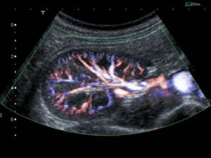
Doppler ultrasound of renal vessels is an additional method of investigation of the urinary system resorted to in order to learn about the condition of the blood vessels that supply blood to the body.
Doppler ultrasound gives information about the diameter of veins and arteries, their location within the kidney, and also allows you to extinct the flow rate of blood.
The principle of the survey is based on the ability of red blood cells reflect the ultrasound waves. A sensor that perceives the answer, transforms it into electrical impulses and displays the finished image.
What are Doppler ultrasound of the kidneys
On the monitor you can see the veins and arteries from the inside, without being invasive (penetrating) intervention in the body. There is an opportunity to recognize the change in blood flow due to spasm of the vessels, the appearance of thrombus or stenosis of the walls (narrowing).
Doppler ultrasound of renal vessels – this is the kind of research that helps to identify the following pathologies:
- a decrease in supply on the blood;
- stenotic changes that lead to narrowing of the arteries or veins;
- vascular disorders that occur due to atherosclerotic changes;
- pathology of the velocity of the blood.
When can an appointment
Not always need to resort to Doppler ultrasound, but the experts recommend to carry out the survey under the following complaints:
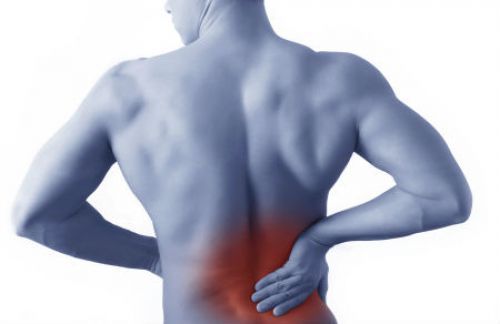
- severe pain in the lumbar region on either side of the spine;
- hypertension without established causes (high blood pressure);
- fall back, which could cause injury or rupture of the kidney;
- cardiovascular disease, which are accompanied by severe edema;
- the development of late toxemia in pregnancy (preeclampsia or eclampsia);
- the presence of renal colic.
Loin – the area of localization of pain in the kidneys

A study of renal vessels can conduct to control the state of the organ after transplantation, or for diagnosis when conventional ultrasound proved to be ineffective.

Important role this test plays in the diagnosis of pathologies in small children. This is because it allows to identify reflux (ing urine from the bladder up), or any congenital abnormalities of the vessels.
Training
The procedure requires careful preparation. It is from the preparatory phase will depend on the quality of the results obtained, the clarity of the picture.

Below the image is more qualitative, a few days before the intended date of survey carried out two preparatory activities:
- The complete exclusion from the diet any foods that promote gas formation in the intestine or slow the process of excretion. These include legumes, bread (especially rye), dairy products, carbonated beverages (sugar water, kvas, beer) and sauerkraut. It is also desirable harder to limit the use of any fruit and vegetables raw.
- 2-3 days before Doppler ultrasound need to start taking enterosorbents is enterosgel, sorbex, and others. Usually appoint 1-2 capsules twice or thrice a day. This measure will help to get rid of excess gases.
Sorbents eliminate intestinal gases
But it is important to remember that you should not change diet and take the sorbents people who suffer diseases such as diabetes, hypertension, coronary artery disease. Since these pathologies require constant medication, and the sorbents do not give active ingredients to be absorbed. In addition, in some diseases contraindicated change diet.
Usually Doppler ultrasound of renal vessels is carried out in the morning and on an empty stomach. In the case where the procedure for some reason was postponed until the evening, then the patient is allowed to have Breakfast. But food should be easily digestible, it should be little, and also it is impossible to suppose, that it stimulated the process of gassing. It is important to remember that the time interval between the last examination and any meal shall be at least 6 hours.
Any specialist should also know that from the survey will be no good, if it holds after studies bowel endoscopic methods (colonoscopy, sigmoidoscopy, EGD). This is due to the fact that during these procedures in the gastrointestinal tract of plenty of air, so the picture on Doppler ultrasound will be fuzzy.
How do Doppler ultrasound
Doppler ultrasound of renal vessels – is absolutely safe, non-invasive studies of the renal arteries and veins. Once trained, the patient is asked to sit or lie down on a couch to the side. The survey allowed to hold two positions, depending on the choice of physician sonology and the patient.

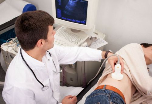
The technique of the procedure is very simple
Then, on the skin in the area of projection of the kidneys is smeared with a special gel lubricant that allows the sensor to closely contact with the body of the subject. The specialist who conducts the procedure, will move the transducer over the skin, thus finding the most successful angle, and the monitor will appear picture in real time.
Doppler ultrasound does not cause any pain and does not last more than an hour. After the examination of patient no restrictions, he can do his stuff.
Standards survey
The doctor-uzist conducts research, and then on a special form writes the conclusion. In it, he indicates these patients and the results of Doppler ultrasound, which characterize the condition of the kidneys and their vessels.
The following is an example of survey results in a healthy person:
- Both kidneys are bean-shaped.
- Their contours are smooth and crisp edges.
- The thickness of the capsule body is 1.5 mm.
- The size of the kidneys are the same (which is allowed the difference does not exceed 2 cm).
- Cups and pelvis is unable to see.
- Echogenicity of kidneys and the liver are the same (there is a variant of the norm when kidney have a slightly lower echogenicity).
- During breathing, the kidneys move no more than 3 cm.
- The resistance index of the main artery is 0.7. In interlobar vessels, it can range from 0.30 to 0.75.
Doppler ultrasound of renal vessels is almost universal method that allows to detect most abnormalities of the body. In addition, it requires no heavy training is not invasive and is done in a few minutes. Also, it is economically much more accessible to patients than, for example, computed tomography. The method has two huge advantages – it does not cause any complications, and to it there are no contraindications.


Email Accuracy Validation through a premium industry level software process to determine if the emails are valid and accurate. Accuracy of between 95 and 97 Bring down your bounce rate on your mass email clients and decrease penalty’s.Lower the chance of your domain address being flagged as spam.
large wedding box