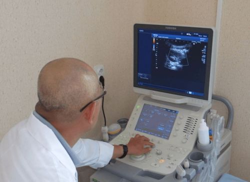
Correct diagnosis involves a full examination of the patient which includes physical, laboratory and instrumental methods. When evaluating the kidneys and abdominal organs most informative method is the ultrasound.
Modern ultrasound is a simple procedure that allows to study the condition of internal organs, determine their sizes, to detect tumors. In order for a qualified expert to be able to qualitatively examine the organs and their supplying blood vessels, the patient must prepare for manipulation.
In this article we will look at what is the proper preparation for an abdominal ultrasound and kidneys and how the procedure is performed.
Why the need for ultrasonic examination of internal organs?
In order to prevent various diseases practitioners suggest patients to undergo instrumental examination of the abdomen and kidneys once a year. This will allow you to monitor the status of bodies, to identify pathological changes and start a rational treatment at a very early stage of development of the disease.

Medical diagnostic branch equipped with modern equipment, through which it expands opportunities of quality and informative examination of abdominal cavity organs and kidneys.
During carrying out of ultrasonic diagnostics of the patients can be identified:
- concretions (stones, sand), which are localized in the bladder, kidneys, bile ducts and gallbladder;
- benign changes of the structural elements of tissues;
- malignant neoplasms;
- the inflammatory processes.
You need to get an ultrasound at the first signs of development of pathological conditions, such as:
- the sharp decline of body weight;
- continuous or periodic nausea;
- disorders of the chair;
- vomiting;
- bleeding;
- the increase of the abdomen;
- acute or chronic pain;
- injury of the peritoneum.
Everyone should know that for the timely identification of problems and stopping its further development with the appearance of disease symptoms should seek professional medical help!
Why the need for a preparation procedure and instrumental studies?
Method of ultrasound has many advantages over other diagnostic methods, such as:
- highly informative – the result of the examination in detail shows the features of the morphological structure;
- time saving – the procedure takes less than an hour, including a description of the outcome of the study;
- painless method eliminates the penetration into the body;
- the safety equipment does not emit hazardous to human body wave.
However, there are many different factors that can influence the outcome of the survey. The main of them is improper training. In such cases, there is dissipation of ultrasonic waves, the distortion of the image. Doctor-sonolog can not accurately identify the pathology, and research data become uninformative. That is why to perform ultrasound examination of abdominal cavity and retroperitoneal space requires careful preliminary preparation of the patient.
Features diet
Before the study of the peritoneum, a person must change your diet at least five days before the procedure. For a detailed examination of the internal organs required to reduce the volume of generated gas in the intestines. Diet before abdominal ultrasound require a complete elimination of that food that causes flatulence (gas accumulation) – raw vegetables, black bread, legumes, fruit, dairy products, juices, coffee.
Flatulence can spoil the picture and complicate the examination of the internal organs
Many patients ask the same question: “How to eat to properly prepare for the ultrasound?”
To obtain reliable survey data, experts recommend to strictly observe the following diet:
- It is important to exclude alcoholic and carbonated drinks.
- People need to eat frequent, small meals daily diet should consist of 5 meals. Allowed to eat: lean boiled or roasted meat; eggs; low-fat boiled or steamed fish; thermally processed vegetables; oatmeal; buckwheat; barley; low fat hard cheese varieties.
- The volume of fluids you drink should be no less than two liters, however during meals water can not drink. Unsweetened tea is allowed an hour after a meal.
The patient will also need to:
- to minimize Smoking;
- to avoid physical and emotional overload;
- to stop taking drugs if this is not possible, it is necessary to warn the physician conducting the examination.
Studies make in the morning, so food it is necessary to refrain for 12 hours. If such prolonged fasting is prohibited for medical reasons (e.g. diabetes), you can drink unsweetened tea and diet crackers.
Other features of the preparatory process
Preparation for an ultrasound includes cleansing the bowel. The day before conducting the study can be mechanical bowel cleansing. For these purposes Esmarch bowl filled with 1.5 l of water. After enema take drugs sorbents – activated carbon, POLYSORB, Enterosgel, Corbatas.

The alternative to this method are:
- the application of therapeutic teas from lemon balm, chamomile, ginger improves digestion and reduces flatulence;
- use laxative decoctions of herbs – buckthorn bark, Senna leaf;
- laxatives – Duphalac, Guttalaks, Sensafirm;
- the use of funds, reduce flatulence – Espumizan, Smecta.
Improves processes of digestion and excretion of harmful substances from the body kidney tea with fennel fruit.
Proper preparation also is to eliminate the patient’s participation in other instrumental studies. Procedures such as rengeo-, gastro – and colonoscopy, MRI with contrast, distort the results of the ultrasound.
In preparation for renal ultrasound specialists recommend to increase the amount of fluid – before the procedure it is necessary to drink about one liter of purified water
Transcript of the study results may be distorted by the following factors:
- overweight patient – a thick subcutaneous fat layer serves as a barrier for the acoustic sensor apparatus;
- skin damage in a zone of examination;
- increased physical activity of the patient (most often this phenomenon can be the child) during the procedure;
- smooth muscle spasm as a consequence of other medical manipulations;
- contrast barium used for other research, can make an image of organs difficult to read.
How to prepare for the ultrasound procedure on children?
In Pediatrics examination using high-frequency waves have been used for over 20 years.

A child is prescribed an ultrasound in cases where there is a need to assess the functional status of the body in:
- congenital abnormalities of the pancreas, spleen, liver, kidneys, adrenal glands;
- disorders of the gallbladder;
- vomiting, frequent regurgitation, low weight gain in newborns;
- the suspected presence of stones in the gallbladder, kidneys, dyskinesia of the bile ducts;
- various injuries of the peritoneum;
- helminthic infection;
- congenital or infectious hepatitis;
- swollen lymph nodes;
- acute pancreatitis;
- anomalies of development of vessels of the abdomen.
Ultrasonic diagnostic method provides a visual picture of all of OBP (abdominal organs), causing the baby absolutely no harm
Preparations of children to study is not much different from training adults. The main objective is to get rid of accumulated gas in the intestines, which is why the kid has to follow a special diet, which excludes milk, pastry sweets, muffins, cabbage, fruit and vegetable juices. In the presence of the child’s disorders of digestion during the four days before the ultrasound, you need to start taking enzymatic drugs that promote quality food processing – Festal, CREON, Mezim.
The study is carried out on an empty stomach, last meal should be no later than 8 hours before the procedure.
Preparation for ultrasound of small children has some peculiarities:
- kids up to 1 year – not to feed for three hours and not drink for an hour before the test;
- children up to the age of three – not to feed for four hours and not drink for an hour;
- children older than three years should not be given food for seven hours, water for an hour before the procedure.
Going on ultrasound, you need to wear comfortable clothing so that the specialist could easily get to the abdomen. To improve the sliding of the sensor on the skin a special gel does not cause allergic reactions. During the procedure, the doctor may ask you to lie on the side, hold your breath, take a deep breath. This is a normal phenomenon that will help you get a clear visual picture of the internal organs. If the patient previously conducted an abdominal ultrasound, you must bring the previous results to diagnostician was able to study the dynamics of changes.