
Method intravenous urography introduced in medicine in 1929, since then the procedure has not lost its diagnostic value and is performed in any medical institution.
The study also called excretory urography that captures the essence of what is happening – excretion (a) contrast material through the organs of the urinary system.
Intravenous urography – a method of radiological diagnosis. It is based on the use of special contrast (iodinated), enter the examinee to a patient intravenously, due to which there is the possibility of visualization of the kidneys and other structures of the urinary tract.
Doing this study all patients who have the outcome of the review of the picture, there are certain violations. The method has certain advantages over such procedures as retrograde pielografia, in particular, it lacks a number of side effects and is suitable for most of the studied patients.
Diagnostic procedures and indications for its implementation
Intravenous urography allows to determine the functional operation of the Cup-pelvis-plating apparatus of the kidneys, ureters, bladder. The accumulation of contrast medium there is a possibility of morphological assessment of these structures.

Iodine is able to very quickly penetrate into the tubules of both kidneys continue unchanged sequentially excreted from the body with the urinary sediment.
The advantage of intravenous urography is the ability to simultaneously visualize the structures of both kidneys, ureters and bladder lumen.
In order to fully visualize all structures need a certain period of time
The method has the following features:
- gives a fairly complete morphological picture of the pathological processes occurring in the cups and pelvis and other structures of the urinary system;
- with sufficient accumulation of contrast agent, it is possible to assess the functional activities of these organs in various diseases (pyelonephritis, renal tuberculosis, traumatic injuries, etc.);
- allows visualization of pathological lesions, stones, foreign bodies and other entities;
- intravenous urography is indispensable for the examination of children, as it allows to avoid such a painful procedure, as the ascending urography, which often have to perform under General anesthesia.
Aiming at study of the child, the doctor saves him from a number of painful or costly procedures (such as cystoscopy or ascending urography)
With proper preparation of the patient to the study, it helps in the diagnosis of the following pathological processes:
- Urolithiasis (formation of stones) at different levels of the urinary tract.
- Hydronephrosis of one or both kidneys.
- Kidney injury, the damage to the ureters or bladder.
- Tumours benign or malignant nature.
- Diverticula in the lumen of the bladder or foreign bodies.
- Dyskinesia of the urinary tract (impaired emptying).
- Tuberculous processes in the kidney.
- Congenital anomalies in the development of organs (e.g., doubling of the kidneys, the ureter bends, etc.).
Special preparation of the patient
In the basis of preparation of the patient for intravenous urography is cleaning his colon from the stool, which can complicate the diagnostic process or to give the doctor a misleading picture of what is happening. It is also necessary to prevent flatulence in the lumen of intestinal loops in the time of the study.
Therefore preparation for intravenous urography starts with assigning the patient a proper diet, which he must adhere for several days before an upcoming study.
The basic principles of nutrition are:
- within 2-3 days before the procedure not used in food legumes and highly starchy foods, eliminates cabbage, fruits and vegetables, you cannot eat white bread and alcohol;
- the evening before the examination the patient is having dinner 17.00-18.00, with the need to give preference to light products;
- on the eve before bed and the next morning you need to spend klishirovannyh “to clean water” or to take special laxatives, which have a mild laxative effect;
- the day before the procedure the patient should limit yourself to a liquid that increases the concentration of the urinary sediment, and therefore improve the process of visualizing the urinary system;
- Breakfast in the morning, better in small sandwich, the liquid can not drink or consume it in very small volume.

In modern medicine, in place of the classic enemas come microclysters, which is very easy to use and does not require the patient’s special skills, acquire them at any drugstore
If the patient needs emergency conduct intravenous urography, when admitted to hospital, he’s getting an enema, and once your bowels start to study.
The algorithm of the study
It must be remembered that before resorting to the procedure and enter the patient iodine-containing drug, the doctor mandatory collect allergic anamnesis. In addition to finding the reactions of allergic character on known patient products and substances the night before, done a skin test using iodine or intravenous administration of 2-3 ml.
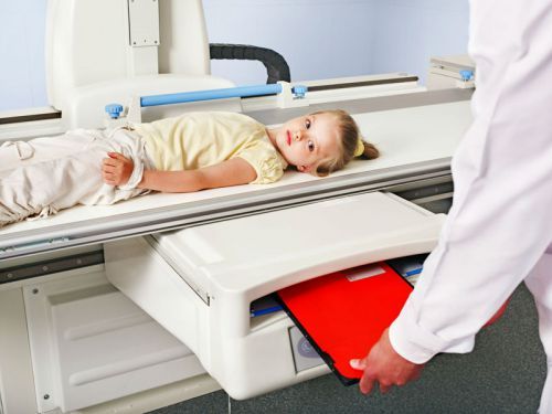
The study is carried out in a special x-ray room, which is equipped with all necessary equipment. The patient is positioned on a special table or couch, and then he is injected 20-30 ml of contrast agent. The introduction is carried out in one of the peripheral veins at the elbow bend.

The substance must enter the bloodstream slowly (injected contrast 2-3 minutes), during this time the patient’s condition should be monitored carefully. Special attention is given to patients of older age groups, patients with the atherosclerotic process in different locations and diseases of the cardiovascular system.
Slow injection of contrast helps to avoid some complications, e.g., anaphylactic shock
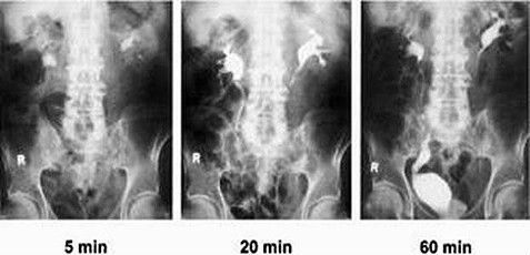
The first series of images is carried out in 5-6 minutes after the contrast has entered the bloodstream. If there is a decrease in functional kidney, this time increases to 10-15 minutes.
If the resulting image does not render the calyx or pelvis on one side, the second study carried out in 50-60 minutes.
Throughout treatments next to the patient must attend the doctor who oversees the entire process and in the early stages of document images (the value and size of the kidneys, their situation, the presence of pathological shadows, etc.).
Contraindications
The method, like any diagnostic procedure, in addition to its advantages, has certain limitations.
Intravenous urography is not carried out, the following categories of patients:
- A serious disease of the kidney, which greatly disturbed their excretory function (introduction of a contrast agent will lead to the deterioration of health).
- Shock the patient’s condition or failure of varying severity.
- Decompensated liver disease, cardiovascular system or lungs.
- Installed an allergic reaction to iodine-containing components.
- Pregnancy (have negative effect x-rays on the body of a child).
- Patients suffering from radiation sickness.
The main advantage of this method is its low invasiveness and relatively high information content. The procedure does not cause the patient pain and rarely accompanied by complications, so still remains the standard of choice in patients suffering from different kinds of diseases in the organs of the urinary tract.


I truly enjoy looking through on this site, it holds great articles.