The unique ability of radioactive isotopes, decaying, radiate energy, which translates to the computer screen and appears as a luminous body that is the basis of this method.
By introducing the isotope investigated the amount absorbed by the kidneys of substances and the rate of filtration of radioactive isotopes.
The development of the science of the 21st century, allows to introduce into medical practice more sophisticated and accurate methods of diagnosis and treatment. One of such methods is radioisotope study of kidney.
In comparison with the method urography radionuclide diagnostics allows you to see the full details about working and the kidneys, to make a series of pictures that represent renografii of the genitourinary system.
What can you learn about the condition of the kidneys?
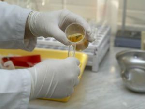
For the conduct of special pharmacological renography uses radioactive drugs that are injected intravenously and once in the kidneys, excreted.
Study allows you to study the function of not only the kidneys but also throughout the genitourinary system, namely:
- the condition of the renal vessels, blood flow, blood supply of the kidneys;
- status and functional ability of the renal parenchyma (renal tissue);
- as the collecting duct system of the renal tubules, pelvis, etc.;
- detects cysts and various neoplasms;
- the state of renal excretory function.
How goes the research?
In the beginning of the study to the patient, as already mentioned, intravenous administration of the special radioactive pharmacological preparation, the radiation of which is designed so that no danger for humans, because the introduction takes into account the body mass. Before the introduction of the drug on the body dress up special sensors that can detect the radiation level. All three of them.
Only after the introduction of a radioactive drug can be conducted further study
After administration to the patient, a series of images that allow to determine the time of promotion of the isotopes of the genitourinary system. All pictures are made in a strict sequence, this is necessary in order to correctly evaluate the function of the receipt of the drug via the bloodstream, and then to evaluate the performance of the collective system, and finally, the function of the excretory system.
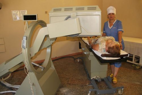
Doing this with a gamma camera in the following way:
- A series of images to determine renal blood flow and renal blood vessels (1 frame per 1 second) is removed within 1-2 minutes.
- A series of photographs to determine the health of renal tissue (1 frame per 1 minute) for 15 – 20 minutes.
- Imagery to determine the collective and excretory functions of the kidneys are conducted after 1-2 hours. They are the finishing.
To conduct radionuclide studies are necessary gamma camera
All manipulations with the patient under a given diagnosis are conducted in the supine position. This study is absolutely harmless and painless.
Indications for
- all chronic inflammatory diseases of the kidneys;
- suspected hydronephrosis kidneys;
- congenital anomalies of the kidneys;
- conditions after kidney injury;
- state after transplantation;
- malignant hypertension;
- suspected growths;
- cystic lesion of the renal parenchyma.
Rules for the conduct of research
In order to properly carry out isotope study of the kidney, the patient has to follow some rules before your procedure:
- stop taking all medications, especially antihypertensive, psychotropic, diuretics;
- the research should be conducted on an empty stomach;
- the day before not to take alcoholic drinks;
- all metal objects are removed;
- the procedure should take place with complete immobility of the patient.
Only observing all these conditions, you can get the right result and to avoid repetition of research.
In some cases, there is a need for re-examination. This happens in situations when you need to track the dynamics of the process occurring in the kidneys or the dynamics and stages of kidney after or during treatment.
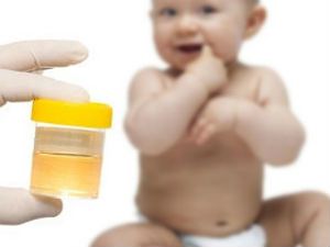
For the radioisotope diagnosis of the kidneys in children there are the same indications as in adults. Usually children younger and middle age the study is not carried out, given that they have unlimited mobility.
Variety of radiological studies
Depending on what type of radionuclide diagnostics need to be applied, are distinguished:
- the radiometry,
- radiography,
- scintigraphy,
- scan.

Radiometry and radiography are methods of diagnosis without the image of the body. Information about the work of the authority is displayed in a graph or chart.
Graphs show the quantification of the kidneys
Scintigraphy, the scan is diagnosis obtain a picture of the organ, and a series of images in layers allows you to see problem areas. Scan on on is special designed for this scanner, approximately 2 hours after drug administration. Images obtained in this way are called “scans”.
Scintigraphy allows to make a series of pictures that have the name “Scintigram”. This data can be played back on the computer screen at any desired time, and considers the necessary areas of the body.
Analysis of the obtained images is performed by a doctor, which is directly specializiruetsya on the radiological studies and interpretation of the obtained images.
Contraindications
To conduct the renography kidney, there is virtually no contraindications. This method of research, as already mentioned, is safe and painless for the patient.
However, conditions such as pregnancy and breast-feeding is the most important and probably the only contraindication for this diagnosis. Children under the age of 1 year the survey is conducted only for health reasons.
A little bit about security
All studies associated with the use of radioactive isotopes, are carried out in the Department of radiology and are perfectly safe for humans.
Drugs are stored in a radioisotope laboratory where in special containers, they are transferred to the office for research. Once opened the vial of isotopes, the remaining dose is placed in a dedicated box for temporary storage. The dosing of the drug the patient is considering his weight, age and pathology severity. All radiological pharmaceutical preparations consist in a special account.
In the radiology Department employs specially trained staff
The exposure during the radionuclide diagnostics is several times smaller than in the x-ray examination. That is why the study can be repeated multiple times if the need arises.
At the end of the business for all specially trained medical staff is monitoring the clothing, hair, hands, shoes. Thus, getting unnecessary radiation to the patients is practically reduced to zero.
In the event of a breakdown, accident or other emergency, Department immediately closes. In order of least penetrating radiation such offices are located in the basement of hospitals. Walls, ceilings in construction are covered by special protective materials. In offices with installed meters are monitoring for the control of radiation. In case of leakage of ionizing radiation the alarm is triggered.
For carrying out radiological examinations of the medical staff have done everything possible to it is held comfortably and securely. If you got a referral to the radiology Department for radiological research, remember that you are assigned to one of the most advanced diagnostic tests in medical practice.