
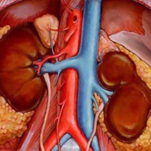
Doppler ultrasound of renal vessels (Doppler ultrasound) is a technique that allows you to explore the particular location of the veins and the arteries concerning the kidneys and also to visualize the localization of the renal vessels, their diameter and structure. During a procedure to evaluate blood flow and identify obstruction in the blood vessels.
High efficiency of the method provides the reflection of ultrasound waves from red blood cells. Ultrasound sensor picks up these waves, converting them into electrical impulses that are displayed on the monitor.
The study is conducted in real time and the image (slice) of blood flow through vessels is displayed on the screen in the form of colour photographs and charts. Examination by ultrasound allows to examine the internal state of the vessels caused by contraction, spasm or thrombosis of unwanted changes.
Order renal ultrasound with Doppler showed the most accurate result, necessary preparation for the study. Adherence to medical recommendations helps you quickly and objectively assess the condition of the arteries and blood vessels.
Indications for
Modern ultrasound machines equipped with a function of Doppler imaging, which allows to combine the information obtained about the movement of blood through the vessels and the information obtained with conventional ultrasound.

USGD vessels of the kidney:
- for the evaluation of blood flow velocity in penetrating the kidney blood vessels;
- to learn about the early vascular disorders that may be caused by atherosclerotic plaques or blood clots;
- to detect narrowing of the arteries (normal up to 5 mm) are important causes of arterial hypertension;
- to detect dysfunction of the blood supply to the body.
Indications for ultrasound of the renal arteries are:
- system increase blood pressure;
- pain in the lower back;
- organ transplant;
- swelling of the legs and puffiness of the face;
- bad results of urine;
- renal colic;
- routine inspection.
The advantages and disadvantages of the procedure
Ultrasound dopplerography is the method of determining blood flow in medium and large vessels, do not only to detect vascular pathological changes, but also to assess the effectiveness of treatment and to identify indications for surgery on the organ.

Doppler is associated with many advantages:
- ultrasound examination of the kidneys is not accompanied by pain because the procedure is minimally invasive (does not involve use of needles or injections);
- for visualization of blood vessels does not apply ionizing radiation;
- Doppler ultrasound has no contraindications and age restrictions;
- unlike x-rays, ultrasound images give a clear picture about the state of the soft tissues;
- the survey is conducted in real time;
- the procedure is the same cost and is widely available.
The main disadvantage is the inability qualitative studies of small blood vessels
Ultrasonic waves can not replace the study of the functional state of the vessels and characteristics of blood flow using radiographic contrast studies – angiography. To clarify the diagnosis after the ultrasound you may need a more thorough examination using a magnetic resonance imaging or computer.
Features of preparation for an ultrasound
An important factor in the effective conduct Doppler ultrasound of renal vessels is the correct preparation of the study. Intestine patient should not contain a large gas accumulation, because such a condition makes it difficult to visualize. In order to reduce gas formation to a minimum, it is recommended prior diet over several days. Such a measure is necessary to obtain the correct result of the procedure.
Usually, patients are recommended to eliminate from the diet foods that promote fermentation in the gut.
Such products include:
- legumes (peas, beans);
- milk;
- freshly squeezed juices;
- black bread;
- strong meat broths;
- raw vegetables and fruits;
- sauerkraut;
- confectionery.
To improve digestion for 2-3 days before the test vessels it is advisable to take “zantac” or “Pancreatin”. Get rid of gases will help such drugs as “Espumizan”, “Activated carbon” or other chelators.
Immediately before the procedure do not drink more than 100 ml of liquid and take drugs diuretic action
Specialists recommend Doppler ultrasound of renal arteries on an empty stomach, so it is best to explore in the morning. If a clinic visit is only possible during the second half of the day, to abstain from food for 6 hours. The recommendations do not apply to people who have disorders that require special diets. For example, this category includes patients with diabetes or taking medications people
The stages of the study
Regular examination of kidneys by ultrasonic wave takes a little time (about 5 minutes). To conduct Doppler ultrasound required a little more time (depending on the nature of the pathology from 10 to 30 minutes).
Doppler ultrasound is not appointed immediately after a colonoscopy or examination of the gastric mucosa. After these procedures, the intestines accumulates air that prevent high-quality imaging of renal blood vessels and arteries.
Ultrasound of renal vessels – informative procedure, but the influence of some factors such as obesity or a tendency to flatulence, adversely affects the reliability of the result. Patients suffering from such problems, examined with breath-hold at maximum exhalation.
Doppler is a doctor-analogom. The patient should sit or lie on your side. Ultrasound is accompanied by the application of a special transparent gel that eliminates an air layer between the device and the patient’s skin.
The gel is washed off easily from clothes and skin, and is Hypo-allergenic, so no irritation. The results of the survey, as a rule, handed out 15 minutes after completion of the procedure.
Decoding the received data
The attending physician, after receiving the results of the diagnosis, assesses the condition of the patient’s body and selects the appropriate treatment.
The conclusion of sonology who carried out the ultrasound must include a transcript, comprising:
- information on the size of the left and right kidneys and their position mobility during respiration;
- information about the edges and definition of the outer contour and the shape of the body (OK – bobulova) and renal cortex;
- data on the coincidence of ehoplotnosti perirenal tissue and renal sinus, also about ehoplotnosti pyramids and parenchyma,
- indicators of the resistance index of the main artery;
- information about visualizing the Cup-pelvis-plating system.
Preparation for ultrasound of blood vessels is very easy, however, following the recommendations, you can help to make the procedure as informative. The diagnostic value of Doppler ultrasound depends on the professionalism of the doctor (sonology), as well as the quality of the equipment. It is therefore advisable to choose a trusted, equipped with good technique clinic with qualified doctors.


Found this on yahoo and I’m happy I did. Well written website.