Scientists are developing custom 3D-printed replicas of human hearts – in an effort to improve life-saving technology fitted in thousands of patients each year.
The models mimic blood flow and pressure in patients’ diseased hearts, allowing surgeons to improve replacement valve procedures and select the best fit.
Replacement heart valves are used when a person’s natural valve is diseased or damaged, stopping the heart from working as it should.
Avoiding potential leakage and failure could remove the need for patients to have multiple risky heart surgeries in their lifetimes.
The prosthetic heart valve market was worth almost $7 billion in 2021 but is expected to more than double in the next decade as the population ages.

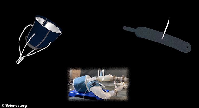
Scientists at the Massachusetts Institute of Technology have created a 3D-printed heart that they can get to pump like a real organ. Pictured above is the heart being developed on a computer
Scientists said the heart could be used to test how valves would work which are implanted into patients suffering from aortic stenosis
This is only the latest medical advance achieved through 3D printing. In December, scientists at the National Eye Institute in Maryland used the technique and stem cells to generate eye tissue.
Experts at the Massachusetts Institute of Technology (MIT), the Cleveland Clinic and other institutions developed the artificial hearts using cans from 15 patients with aortic stenosis, a narrowing of heart valves that impedes blood flow.
Everyone’s heart contains four valves that stop blood from flowing backward with each beat, allowing for a steady supply of oxygen and nutrients.
But in aortic stenosis, the valve between the lower left chamber — ventricle — and the main artery — aorta — becomes narrowed and doesn’t fully open.
This leaves the left ventricle needing to work harder, causing chest pain, and may also cause oxygen-rich blood to start to flow backward into the lungs.
About 1.5million people in the United States suffer from aortic stenosis, with four-fifths of those who don’t get treatment dying within five years.
Patients currently receive two different valves: Either the self-expanding Evolut R — which enlarges to fill the gap where the valve is when it is heated — or the SAPIEN 3 — which is put in place of the damaged valve using a balloon.
Medics at MIT hope that their replica heart could be made for each patient to test which valve would work best.
It could also be used to help in the treatment of other patients, they said, such as those with congenital heart disease, one or more problems with the heart’s structure that exist since birth.
Similarly, the device could be used in industry to help to develop new treatments and in colleges to train future doctors.
Dr Luca Rosalia, a graduate student in the MIT-Harvard Program, told DailyMail.com the replica takes about a day to make. He was not clear on the costs.
The model, revealed this week in Science Robotics, was developed using images from 15 patients with aortic stenosis.
Images were converted into a three-dimensional computer model of their left ventricle and aorta using polymer ink.
Elastic sheets were also applied to allow the replica to expand and contract, just like a real heart, and a rigid skeleton was applied to ensure that it kept its shape.
Scientists also used the machines to print a 3D model of the patient’s aortas. This model included the valve that was narrowed or partially closed.
Once created, the heart was connected to a system that pumps air in and out, mimicking the flow of blood in a patient with aortic stenosis.
They then tested applying different valves to the model, to measure how the flow of air — a substitute for blood — changed.
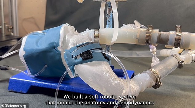
This image shows the model with tubes attached to pump air in and out – which mimics blood. The doctors said it began to work just like a real heart. The model is of a left ventricle and aorta
Pictured above is the model heart being held by one of the researchers
Asked whether this model brought the scientists closer to making a 3D heart that could be transplanted into patients, Mr Rosalia told DailyMail.com that was the case.
‘Some of the soft robotic sleeve elements could be used to support cardiac function in patients with aortic stenosis,’ he added. ‘[But] our work is preclinical.’
‘This is a model, but we envision it being built for every patient that comes in’ to test the impact of replacing the aortic valve in the heart.
‘There is a big need to have a model that is patient-specific and can also simulate cases where there is an anomaly in the morphology of the aortic valve,’ he said.
‘We have patients who as they get old, it gets narrower, and others who are born with congenital defects [who could benefit].’
Co-author Dr Ellen Roche, of Massachusetts General Hospital, said: ‘Being able to match the patients’ flows and pressures was very encouraging.
‘We’re not only printing the heart’s anatomy but also replicating its mechanics and physiology. That’s the part we get excited about.’
When valves were implanted as they pumped the researchers observed they produced similarly improved flows as seen in patients following their procedures.
Co-author Dr Christopher Nguyen, of the Cleveland Clinic in Ohio, said: ‘Patients would get their imaging is done, which they do anyway, and we would use that to make this system – ideally within the day.
‘Once it’s up and running clinicians could test different valve types and sizes and see which works best – then use that to implant.’
Warning signs of aortic stenosis
Aortic stenosis happens when the opening of the main valve, which takes freshly oxygenated blood from the heart to the rest of the body, narrows, reducing the blood flow.
The disease is usually caused by a build-up of calcium – a mineral in the blood – on the valve.
This increases naturally with age but smoking, high blood pressure and obesity can accelerate it.
The heart must then work harder to pump blood around the body, which in turn receives less oxygenated blood.
Often the first signs of damage to the aortic valve go unnoticed. Sufferers usually live unaffected for several years before symptoms start and they seek help for treatment.
Symptoms of aortic valve stenosis often only appear when it is advanced. These can include:
- Breathlessness
- Chest pain
- Fluttering in the heart
- Feeling faint or dizzy