
Magnetic resonance imaging is the most informative, accessible and safest method of inspection of organs of the urogenital system. Diagnostics allows to obtain clear, three-dimensional images of the examined region of the patient.
Detect pathology of the pelvic organs, and in some cases, and rectum in women and men in the early stages of development, and also to control the dynamics of patient’s status and to make timely changes in the treatment plan.
The accuracy and information content of images is largely dependent on proper preparation for MRI of the pelvis.
How is magnetic resonance imaging
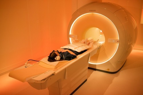
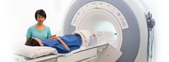
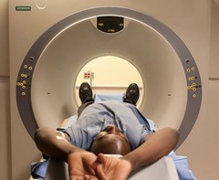
The MRI apparatus of the closed type made in the form of a cylindrical tube surrounded by a magnet. The patient lies on a movable part of the equipment – table and automatically moves to the interior of the device.
The use of diagnostic equipment open type allowed for examination of patients with claustrophobia, and overweight.
Maximum efficiency high-definition images are the open machines with high magnetic field of over one Tesla.
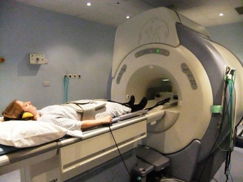
For examination of pelvic organs magnetic coil is placed on the pelvic area and the patient for half an hour needs to maintain the stationary state. If the diagnosis is carried out using a contrast agent, its duration increases to one hour.
Note: the procedure is painless, but some patients experience discomfort caused by the magnetic field. The patient is a hardware one, but between him and the doctor established audio, in addition, the specialist sees the patient and supervises the procedure. After the diagnosis will not need time to adapt.
The advantages of magnetic resonance imaging
- Diagnostics noninvasive and is performed without the use of dangerous radiation used in radiology.
- Magnetic resonance imaging allows to obtain the most clear, informative pictures.
- In the course of diagnostics of pathologies can be detected at the initial stage of their development. This applies to malignant neoplasms, injuries and diseases that are asymptomatic.
- The solution used for the procedure, does not contain in its composition of iodine is not an allergen in contrast to iodine-containing substances, the use of which is common in radiography.
- Preparing for MRI of the pelvis does not require that patients perform serious action.
Possible risks in MRI
- Diagnosis safe for the health of the patient, provided careful attention to the recommendations of the expert.
- The magnetic field poses no danger to health, but may provoke the malfunctioning of the implanted devices based on metal.
- Allergic reactions are rare and easily dealt with by light drugs.
Note: in some cases, an MRI is performed in combination with other surveys – ultrasonic inspection, radiography.
Features MRI of the small pelvis in women
The main advantage of the survey is a timely opportunity to see the changes of a pathological nature at the cellular level.
Experts do not recommend routine screening to women in the following cases:
- The first trimester of pregnancy. Magnetic hollow may adversely affect fetal development, so the possible fact of pregnancy must inform the specialist.
- Of renal disease and diabetes. In this case, it is not recommended to use a contrast agent, since its components can cause disturbances in the functioning of the kidneys.
- In case of heavy blood discharge during menstruation, in this case to determine pathology in the pelvis impossible.
- If the patient is a nursing mother – after the diagnosis should not feed the baby for two days.
- In the presence of intrauterine devices made of copper screening, as a rule, not carried out, since under the influence of the apparatus it can shift.
As a woman to prepare for MRI of pelvic organs
Special training is not necessary, but to implement some of the recommendations is necessary, otherwise the survey will lose the information content, and the results will be distorted.
So, before conducting diagnostics should consider the following factors:
- Menstrual cycle. The optimal period for survey – on the seventh day of the cycle.
- Diet. For several days before diagnosis should be excluded from the menu of beverages with high gas content, fiber and dairy products.
- An hour before the examination need to visit the toilet and to drink activated charcoal (for every 10 kg of body weight have two tablets of the drug).
- The last meal must be six hours before the test.
Note: the below procedure was completed efficiently and the results reflect the status of the health of the patient, shall provide to the specialist medical history, including previous surveys.
Features MRI of the pelvis in men
Diagnosis in men, primarily aimed at identifying damage to the pelvic organs, inflammatory processes. In urology, MRI allows us to diagnose malignancy in the urethra or thrombosis.
The procedure is performed at frequent filling of the bladder, with restrictions in the diet are not provided. The survey duration varies from 10 minutes to an hour.
If the patient is difficult to keep still, he needs to be sedated in some cases anesthesia is used.
The survey is not carried out for patients who have a pacemaker, insulin pump, metal braces and even dentures made of metal. Also an obstacle to the procedure is allergic to the active substance of the contrast solution. If claustrophobia is assigned to another kind of diagnosis is less dangerous for the psyche of the patient.
Algorithm for MRI using contrast solution similar to the normal procedure of examination. The only difference is that diagnosis begins immediately after the introduction of the solution.
Note: magnetic resonance imaging is used in the examination of the genitals and scrotum, such diagnostics are especially needed in sports because of frequent injuries.
Preparation for the MRI study of the pelvis in men
Preparations for the MRI takes much less time and easier in comparison with a preparatory stage for other types of surveys. Not require clinical training, as well as medication. Enough to stick to a light diet during the day before diagnosis, which is the limited use of products containing gases, lactic acid bacteria and fiber. You should also avoid eating spicy seasonings. The last meal should be three to four hours before the procedure. Preferably immediately before the examination to drink some tablets “But-shpy” or “Espumizan”.
If you plan to use a contrast agent, before the need to do an enema. This type of survey is only administered in cases of suspected malignant tumors in the prostate gland, the lymphatic system and the urinary tract.
It is mandatory to analyze for allergic reactions to substances with a contrast solution.
To determine the diagnosis and administration of appropriate regimens, it is important to decipher the images. First image is studied by the specialist who conducted the survey, gives a preliminary conclusion. The final findings and treatment the patient receives from a physician.

