Often diagnostics of the oral cavity requires not just a single shot of the tooth, but the full picture. In such a situation the most appropriate solution is a panoramic image of teeth, or orthopantomogram. Sometimes it is the only method that allows you to put the correct diagnosis.
HOW IS THE PROCEDURE
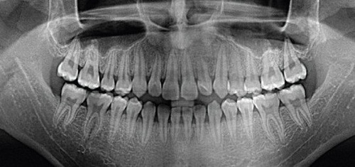
Orthopantomogram (abbreviated as OPG) is a circular panoramic image of teeth in which the jaw is entirely deployed in the plane. In addition to the teeth, roots canals, crowns and fillings, in this photo you can examine the state of periodontal tissue, maxillary sinuses, sinuses.
Panoramic picture of the teeth also helps to detect hidden cavities, cysts, inflammation, periodontal pockets, abscesses, to perform the exact condition of the roots and periodontal tissues, temporomandibular joints, maxillary sinuses. In addition, OPG is indispensable if you plan an orthodontic treatment with aligners or braces.
In preparation for the implantation of the teeth in the panoramic shot, the doctor can determine whether enough bone tissue for implantation or required sinus augmentation how many implants are required and what their optimal size and what will be the dynamics of their healing.
It is extremely important to make a panoramic overview, to determine the position and condition of wisdom teeth. That’s why competent doctor decides to remove or cure the “eight” only after the analysis of the panoramic image.
HOW IS A PANORAMIC IMAGE OF TEETH
No preparation for orthopantomogram not needed. It is sufficient to only remove metal jewelry – necklaces, earrings, piercing to the picture without distortion and unnecessary artifact did not cover the separate areas of the image.
With the help of a technician the patient takes place in the unit, wearing a protective apron and collar, then bites down on a special plastic holder. Then an x-ray emitter rotates around the patient’s head.
DIGITAL OR FILM PANORAMIC RADIOGRAPHY: WHAT TO CHOOSE?
Digital equipment today is almost ousted from the practice film counterparts. Although they cost the clinics more expensive, but then do not require expenses for supplies and equipment photo lab.
In addition, tape devices have greater degree of exposure.
A panoramic image taken by a digital method, is instantly transferred to the computer doctor and is entered into an electronic patient chart. If desired, the latter may obtain the on disk or in printed form.
Finally, the image quality in the case of digital equipment is much higher than with film. So the expert receives better information to work with.
THREE-DIMENSIONAL ORTHOPANTOMOGRAM
Panoramic image of the jaw in 3D is performed on the computer tomograph. This unit captures the study area in different projections, and then creates a three-dimensional image. This allows the doctor most effectively to study the structure of the jaw for the diagnosis and subsequent treatment.
Today orthopantomogram are available in any dental clinic.