
The most emergent diagnosis of consideration, a septic joint, occurs less frequently in the elbow than in larger joints (5-10%), with the exception of cases in which Neisseria gonorrhoeae is the pathogen.2, 3 For more information, see Surgical Treatment of Septic Arthritis.
Background
Arthrocentesis involves both the puncture of a joint and the aspiration of its synovial fluid. It is typically used to make an accurate diagnosis of a painful, warm, swollen joint. Removal of excess fluid can be therapeutic. Analysis of the removed fluid helps to decipher its etiology.1
Indications
Diagnostic
Before the procedure, periarticular processes such as bursitis, tendinitis, contusions, and cellulitis must be excluded on clinical grounds. Performing an arthrocentesis with the goal of obtaining synovial fluid to send for analysis is useful for the following purposes:
-
Evaluation of a nontraumatic acute monoarticular arthritis
- Evaluation of a suspected infection in an elbow (eg, septic arthritis) 4, 5
- Evaluation of a possible inflammatory cause of an effusion (eg, gout, pseudogout, 6 rheumatologic disorders, reactive arthropathies)
- Differentiation of an occult fracture not clearly visualized on radiographs from an inflammatory cause by the presence of an hemarthrosis
To date, the diagnostic value of elbow joint aspiration for periprosthetic elbow infection has not been established.7

Therapeutic
A large effusion caused by fluid or blood is painful and leads to significant impairment of joint mobility.
Removal of synovial fluid as a therapeutic modality is useful for the following purposes:
- Relief of pain
- Improvement of mobility
- Instillation of medications
- Repeated arthrocentesis for a septic elbow in carefully selected patients as a means of decreasing bacterial load and avoiding surgical debridement (only under the discretion of the orthopedic specialist)
Elbow arthrocentesis has been employed as an initial treatment measure in patients with radial head fractures; however, a 2014 Cochrane review did not find sufficient evidence to permit determination of the effectiveness or safety of this procedure in adults.8 A subsequent systematic review by de Muinck Keizer et al found that in patients with Mason type I radial head fractures, aspiration of hematoma appeared to be safe and effective.9
Contraindications
Arthrocentesis through overlying cellulitis is controversial because of concerns about seeding the joint with bacteria. This decision should be made in consultation with an orthopedist and with consideration of the potential risks and benefits.
Relative contraindications for elbow arthrocentesis include the following10 :
-
Overlying skin or soft-tissue infection (eg, cellulitis or abscess; see above)
- Overlying skin lesions (eg, dermatitis or psoriasis)
- Known bacteremia
- Bleeding diatheses
- Patient on anticoagulation medication
- Prosthetic joint (refer to orthopedic specialist)
Periprocedural Care
Equipment
Equipment used for elbow arthrocentesis includes the following:
-
Sterile gloves and towels
- Skin cleanser (povidone-iodine, chlorhexidine)
- Local anesthetic agent (lidocaine 1%, vapor coolant)
- Small (25 gauge) needle and small (5 mL) syringe for anesthetic injection
- Needle (18-20 gauge) and syringe (5 or 10 mL) for joint aspiration
- Specimen tubes (ethylenediaminetetraacetic acid EDTA for cell count and differential, lithium heparin for crystals, and a culture tube)
- Sterile gauze
- Elastic wrap, if needed
Patient Preparation
The synovial membrane contains pain fibers; therefore, customary practice is to instill a local anesthetic before the procedure to minimize pain. (See Local Anesthetic Agents, Infiltrative Administration.) Typically, lidocaine 1% is used. After the skin is prepared with a povidone-iodine solution or chlorhexidine, make a small wheal with a small (25-gauge) needle in the dermis at the determined entry point for aspiration. Do not inject anesthetic into the joint, because this may hinder synovial fluid analysis.
Alternatively, a topical vapor coolant, such as ethyl chloride, may be sprayed on the skin before needle aspiration. Infrequently, procedural sedation may be required in young children or uncooperative patients.
Positioning
Place the patient sitting upright on a stretcher. Bend the patient’s elbow to 90º. Pronate the patient’s forearm, and rest it with the palm down on a side table set at the appropriate height for comfort.
Technique
Aspiration of Synovial Fluid From Elbow
Explain the procedure to the patient and obtain informed consent.
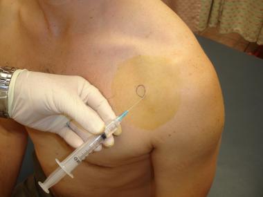
With the patient positioned as described (see Positioning), identify the olecranon process, lateral epicondyle, and radial head, and find the depression (or bulge, if the effusion is large) in the soft triangle. (See the image below.) This site is used for all approaches.11 The landmarks may be easier to find if the arm is first extended to locate the depression and then flexed and pronated for the procedure. Do not confuse an olecranon bursitis with a joint effusion; an olecranon bursitis is located posteriorly over the olecranon.
Triangle formed between olecranon, lateral epicondyle, and radial head as site for needle placement.
Identify the site of entry, and mark the site with a plastic needle sheath or sterile marker. Prepare the skin with a cleansing agent, and drape with towels.
Do not pass the needle through a site with cutaneous signs of infection unless the source is strongly suspected to be from the joint. This should only be done after consultation with an orthopedist.
Anesthetize the area by injecting 1-2 mL of lidocaine 1% and forming a skin wheal. Insert an 18-gauge needle into the depression perpendicular to both the skin and radial head from the lateral side. This is the lateral approach, which is preferred. A medial approach should not be used, because of the risk of injury to the ulnar nerve and the superior ulnar collateral artery.
As an alternative to the lateral approach, the posterolateral approach can be used. An increased risk of injury to the radial nerve and triceps tendon exists, but this approach is useful if the bulge of an effusion is palpated inferior to the lateral epicondyle. In the posterolateral approach, insert the needle perpendicular to the skin but parallel to the radial shaft. (See the image below.)
Aspiration of elbow via posterolateral approach.
Ultrasonography may aid detection of even a small effusion in the olecranon fossa.12, 13, 14
Advance the needle slowly while aspirating the syringe until synovial fluid is obtained. If the aspiration is unsuccessful, draw back, reidentify the landmarks, and correct the needle insertion position. If bone is encountered, withdraw the needle slightly and redirect it.
When synovial fluid is obtained, remove the needle. Apply a bandage and elastic wrap if a large effusion was present. Place the fluid in specimen tubes and send for analysis.
Complications
rthrocentesis of the elbow or any joint is associated with infrequent complications, including the following:
-
Infection – In rare cases (1 per 10,000 aspirations), bacteria from the skin may be introduced into the joint 2
- Iatrogenic hemarthrosis – A small amount of blood may be obtained from a small synovial blood vessel, which is a benign occurrence; obtaining larger amounts is rare, except in patients who are taking an anticoagulant or who have a history of bleeding diatheses
- Allergic reaction – Hypersensitivity to the anesthetic agent, if present, can be identified from the history
References
- Bettencourt RB, Linder MM. Arthrocentesis and therapeutic joint injection: an overview for the primary care physician. Prim Care. 2010 Dec. 37 (4):691-702, v. Medline.
- Thomsen TW, Setnik GS. Arthrocentesis: elbow (emergency medicine). Procedures Consult. Available at http://www.proceduresconsult.com/medical-procedures/arthrocentesis-elbow-EM-057-procedure.aspx. Accessed: October 19, 2018.
- Polishchuk D, Tan V. Atraumatic septic arthritis of the elbow in a young immunocompetent host secondary to distant hematogenous spread. Am J Orthop (Belle Mead NJ). 2012 Aug. 41 (8):369-70. Medline.
- Lin HM, Learch TJ, White EA, Gottsegen CJ. Emergency joint aspiration: a guide for radiologists on call. Radiographics. 2009 Jul-Aug. 29 (4):1139-58. Medline.
- Nduaguba AM, Flynn JM, Sankar WN. Septic Arthritis of the Elbow in Children: Clinical Presentation and Microbiological Profile. J Pediatr Orthop. 2016 Jan. 36 (1):75-9. Medline.
- Tai CH, Oh HB, Seet JE, Ngiam KY. Pseudogout – a rare manifestation of hungry bone syndrome after focused parathyroidectomy. Ann R Coll Surg Engl. 2018 May. 100 (5):e106-e108. Medline.
- Somerson JS, Morrey ME, Sanchez-Sotelo J, Morrey BF. Diagnosis and Management of Periprosthetic Elbow Infection. J Bone Joint Surg Am. 2015 Dec 2. 97 (23):1962-71. Medline.
- Foocharoen T, Foocharoen C, Laopaiboon M, Tiamklang T. Aspiration of the elbow joint for treating radial head fractures. Cochrane Database Syst Rev. 2014 Nov 22. 11:CD009949. Medline.
- de Muinck Keizer RJ, Walenkamp MM, Goslings JC, Schep NW. Mason Type I Fractures of the Radial Head. Orthopedics. 2015 Dec. 38 (12):e1147-54. Medline.
- Reichman E, Simon RR. Arthrocentesis. Emergency Medicine Procedures: Text and Atlas. New York: McGraw-Hill; 2004. 559-67.
- Sanford SO. Arthrocentesis. Roberts JR, Custalow CB, Thomsen TW, Chanmugam AS, Chudnofsky CR, DeBlieux PMC, et al, eds. Roberts & Hedges’ Clinical Procedures in Emergency Medicine. 7th ed. Philadelphia: Elsevier; 2019. 1105-24.
- Fessell D, van Holsbeeck M. Ultrasound guided musculoskeletal procedures. Ultrasound Clin. 2007 Oct. 2:737-57. Full Text.
- Reijnierse M, Miller TT. Video: Musculoskeletal ultrasound imaging of the elbow: part 2, Pathology. AJR Am J Roentgenol. 2013 Jun. 200 (6):W645. Medline.
- Boniface KS, Ajmera K, Cohen JS, Liu YT, Shokoohi H. Ultrasound-guided arthrocentesis of the elbow: a posterior approach. J Emerg Med. 2013 Nov. 45 (5):698-701. Medline.


I want forgathering useful info, this post has got me even more info! .
Hello there,
My name is George, and I was wondering if you would like to have your website osvilt.com promoted as a resource on my blog
We are updating our broken link resources to include up to date resources for our readers. Our resource links are manually approved as a do follow link.
If you are interested in having your site included as a resource on our blog, please let me know.
Thanks for your consideration,
George
So this post abslutely made me think! TY-I wouldn’t have thought of things this way otherwise.
Thanks for your post. Another element is that being photographer involves not only difficulties in catching award-winning photographs and also hardships in establishing the best digicam suited to your requirements and most especially issues in maintaining the standard of your camera. This really is very correct and evident for those photography addicts that are in capturing this nature’s engaging scenes — the mountains, the forests, the actual wild or even the seas. Going to these amazing places definitely requires a dslr camera that can live up to the wild’s hard natural environment.
I can think of more reasons that support this. Can I share an excerpt?
Hey,
Charles the promotion guy said I should send you this to help with your site on osvilt.com. We discovered this coupon code to get some free content to use on the blog, you know, to keep things fresh and updated to stay on the good side of Google. This is normally $40 worth of goodies!
Best,
Earle
I抦 impressed, I must say. Actually not often do I encounter a blog that抯 both educative and entertaining, and let me tell you, you might have hit the nail on the head. Your concept is outstanding; the problem is something that not enough individuals are talking intelligently about. I’m very completely satisfied that I stumbled throughout this in my search for something relating to this.
There is noticeably a bundle to learn about this. I assume you have made particular nice points in functions also.