Defartrosis is divided into two groups, primary and secondary, depending on the cause of its development.
The primary begins, as a rule, for no apparent reason, its genetic predisposition becomes the dominant factor.
In addition, age-related changes in tissues accompanied by a sedentary lifestyle can also cause the onset of pathology.
The secondary form has visible and deeper causes of the formation of the disease:
- large loads on the joint, more often in athletes or overweight people;
- injuries or consequences of operations;
- anomalies in the development of the musculoskeletal system (static skeletal deformities, dysplasia);
- concomitant endocrine diseases (hormonal disorders, diabetes mellitus, acromegaly);
- circulatory disorders in the joints;
- metabolic changes (chondrocalcinosis, gout, hemochromatosis);
- cartilage inflammatory processes;
- phlebeurysm.
Arthrosis of different localization affects about 8-12% of people in the world, it is more common after forty yearsIn children, pathology can develop as a result of trauma or associated musculoskeletal diseases. Deforming arthrosis of the knee joint (gonarthrosis) is one of the frequent places of the disease’s location, which is characterized by damage to the cartilage with further involvement of the adjacent bone, muscles, ligaments.
Classification of Gonarthrosis
The clinical picture depends on the degree of progression of arthrosis, at first the pathology is accompanied by aching pains that increase with exercise. The discomfort gradually increases, lameness appears, the need for support, closer to the last stage, ascent or descent down the stairs becomes an insurmountable obstacle.

In more detail, the symptoms can be described by the degree of the course of the disease.
- First degree. It is characterized by a slight swelling of the joint associated with the accumulation of fluid, in the case of a large amount of edema, edema may develop, other visible changes are absent.
- Second degree. The manifestations of this stage are more pronounced, swelling becomes stronger, joint deformation occurs, accompanied by pain with minimal physical exertion, a crunch is clearly audible. During palpation, joint expansion is diagnosed, it is difficult for the patient to bend the knee.
- The third degree of the pathology course is considered the most difficult, pains become unbearable even at rest, standing and walking becomes problematic. Joint deformity becomes apparent. The gap between the joints is completely absent, bone growths occur, atrophy of the thigh muscles can occur.
Diagnostics
The examination takes place in several stages:
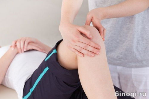
- collection of medical history, examination and palpation of the knee joint;
- radiography, the main examination that helps to accurately establish the diagnosis, the picture shows a decrease in the distance of the joint space, marginal osteophytes may also be observed;
- laboratory research involves a puncture from the gap of the joints, if synovial fluid is detected, this indicates the presence of arthrosis;
- in addition, on the second or third degree of the course of the disease, an MRI of the knee joint may be necessary.
Conservative therapy
Deforming arthrosis of the knee is a chronic and progressive disease, with periodic remissions and relapses. Conservative treatment is possible at the first or second degree of arthrosis, the third requires surgical intervention.
Conservative treatment can not completely eliminate the pathology, but only transfer it to the stage of remission, the main goals of this type of therapy include:
- slowing down the progress of arthrosis;
- improving the quality of life of the patient, by reducing pain;
- partial restoration of joint function.
A comprehensive course of treatment consists of:
- drug therapy;
- physiotherapeutic procedures;
- diet
- medical gymnastics.
Drug therapy
Drugs are prescribed topically, orally, and as an intraarticular injection.
In order to reduce the progress of the disease and reduce pain, non-steroidal, anti-inflammatory drugs are used:
- Diclofenac;
- Naprsen;
- Ibuprofen;
- Voltaren;
- Indomethacin;
- Pyroxycycline;
- Elderin;
- Celebrex;
- Movalis.

The following group of medicines is intended to improve blood circulation and microcirculation:
- Drotaverine;
- Trental;
- Pentilin;
- Curantyl;
- Pyridoxine;
- nicotinic and ascorbic acid.
Derivatives of hyaluronic acid are administered intraarticularly, in extreme cases glucocorticosteroid preparations are prescribed, as a rule, for stopping synovitis.
No more than three such injections are allowed per year with an interval between each of four months. The use of glucocorticosteroids is dangerous because it causes the progression of destruction of the articular cartilage, as well as secondary bone necrosis.
All drugs for treatment are prescribed individually for each patient based on the clinical picture and the degree of progression of arthrosis, self-medication is prohibited.
A group of chondroprotectors improves the elasticity of adjacent tissues, and repair a damaged joint. Greater effectiveness from the use of funds is observed at an early stage of the disease and their long-term use.
Apply:
- Chondroxide (Outer);
- Arthron Hondex (oral);
- Chondroitin Complex (oral).
Physiotherapy
Gymnastics helps strengthen muscles and improve blood circulation in a sore joint. After two to three weeks of systematic training, patients note a general improvement in their condition. It is necessary that the set of exercises was coordinated with the orthopedist, consisted of gentle exercises with minimal physical stress on the joint.
Excessive efforts can lead to the opposite result and accelerate the destruction of cartilage.
All classes should be carried out in a lying or sitting position, so as not to create additional load on a sore leg. Aerobics, swimming, knee massage are welcome, all procedures should be done in a dosed manner.
Therapeutic gymnastics can be used only during the period of remission, during exacerbation the joint needs complete rest.
Physiotherapeutic procedures
The complex treatment must necessarily include physiotherapeutic procedures, they have a positive effect on the sore joint, act as a local analgesic, improve blood circulation and eliminate muscle spasm.
Often prescribed:
- UHF;
- magnetotherapy;
- paraffin and ozokerite applications;
- exposure to ultrasound;
- electrophoresis with novocaine or analgin;
- laser therapy;
- phonophoresis with hydrocortisone;
- inductothermy;
- cryotherapy;
- radon and hydrogen sulfide baths.
Which of the following procedures are suitable for treatment in your case, only a specialist should decide.
Diet food
One of the causes of arthrosis is considered overweight, it is observed in most patients with this disease. Therefore, it is important to follow a diet aimed at the need to reduce body weight. It is noticed that people who have lost at least five extra pounds with a diet noted a general improvement in their condition and a decrease in discomfort in their sore knee, especially while walking. Doctors recommend a diet for all patients with gonarthrosis, regardless of overweight, as certain foods can help restore cartilage and strengthen bones.
Products and dishes, the use of which is necessary during the diet:
- cottage cheese;
- hard cheeses;
- buckwheat;
- lentils
- lean meat and fish;
- jelly from bone broth;
- fish broths;
- dishes with the addition of edible gelatin;
- bananas
- chicken eggs.
During the diet, it is necessary to exclude alcohol and reduce the consumption of fatty foods, tomatoes and sour, citrus fruits. Dishes can be steamed, stewed or boiled, as much as possible to limit frying.
Surgical treatment
If conservative treatment does not bring proper results and the disease progresses, they resort to surgical intervention. Most often, corrective osteotomy is performed, its main goal is to achieve a uniform load on the joint. Corrective osteotomy gives good results, in 80% of patients there is a significant improvement in the condition of the knee, restoration of the joint space and a decrease in pain during an average of ten years.
Osteotomy has its contraindications, these include:
- pathology of the adjacent joint;
- cartilage deformation exceeding 15 °;
- age after 60 years;
- joint instability;
- the inability to bend the knee by more than 90 ° and extension by 15 °.
Before corrective osteotomy, a more gentle procedure can be prescribed – arthroscopy.
In addition to corrective osteotomy, doctors can resort to ventralization of the tibial tuberosity or endoprosthetics. It is worth noting that, compared with osteotomy, arthroplasty is a very complex and expensive operation requiring special implants and suitable equipment. Such an operation is carried out only in specialized orthopedic centers in case of emergency.
Deforming arthrosis of the knee joint, a disease that does not occur suddenly, but develops gradually as a result of many related factors. Of course, the best treatment is prevention, but if arthrosis has already attacked you, you should responsibly take the course of therapy at an early stage of the disease to prevent its progress and subsequent surgical intervention.
Using supportive therapy and following all the recommendations of a doctor, it is quite possible to get rid of uncomfortable feelings by achieving long-term remission and continue to enjoy a full, active life.