
Modern medicine has a large Arsenal of methods of diagnosis of various diseases of the intestines and stomach. Each of them has its advantages and indications for. A generic method that would replace all others, does not exist.
All diagnostic studies in the complex allow you to determine the status and identify any existing pathology.
Common methods of instrumental study is colonoscopy and magnetic resonance imaging. Called diagnostic procedures have advantages over others. However, often patients have a question, what is better – an MRI of the bowel or colonoscopy. The unequivocal answer can not be, because each of these methods has specific indications for and has its own characteristics.
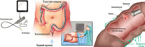
Colonoscopy
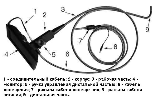
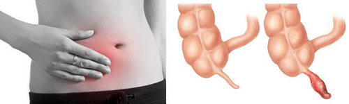
Is one of the most informative among instrumental investigations. It allows you to assess the condition of the body wall and identify any pathological changes. Possible fence cellular material for biopsy and removal of polyps.
The method consists in the introduction through the anus endoscopic probe, which is a video camera that transmits images to the monitor. At the same time in the intestines evenly air is supplied to smoothing that allows for easy advancement of the colonoscope and visualize the entire inner surface of the body.
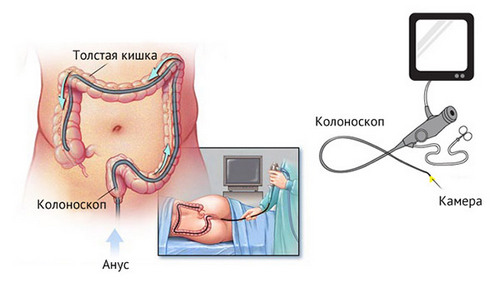
Colonoscopy is considered to be a highly informative method, which allows to accurately study the condition of the large intestine: straight to the cecum. But it has its drawbacks, primarily of physical discomfort in it. It is the fear of discomfort often leads patients to question the necessity of completing this research or even to abandon it.
Actually the procedure is much easier than many imagine. Before the introduction of the endoscope is carried out anesthesia, which makes it possible the pain is minimal. Recommended before colonoscopies receive antispasmodics further facilitates its implementation.
The study runs for 10-30 minutes depending on the required length of the path traveled by colonoscope. After completion of the procedure introduced air is aspirated, and all the discomfort from bloating disappear.
MRI of the colon
This study allows to obtain a three-dimensional image by applying a magnetic field. To scan the abdomen of the patient is placed in a special MRI installation, so that it was in the center of the radiation. For better visualization of blood flow, detect tumors and inflammatory defects of the used contrast agent.
A significant advantage for patients considered painless MRI of the intestine and the lack of need to introduce inside of any tools. That is why most people prefer this kind of diagnosis. Imaging is best done to detect abnormalities of the small intestine and stomach, which are well visualized on the images. In relation to Tolstoy’s Department specified method is designated as optional, because it is not possible to examine the state of the body wall having a plurality of folds from the inside.
Comparison of research methods
Colonoscopy and magnetic resonance imaging are highly informative ways, greatly facilitate the correct diagnosis. Comparing them is difficult because they have different algorithms for the execution of its advantages. The advantages of MRI is the comfort of its implementation, the ability to determine the presence of intestinal obstruction and tumors of the body beyond its limits. In other cases, imaging is inferior to colonoscopy in diagnostic of many aspects.
While MRI is not carried out the collection of material for biopsy and therapeutic measures such as polypectomy (removal of polyps). Colonoscopy produced similar manipulations. The endoscope easily overcomes the bends of the loops and allows to study the state of the body, even in “hard to reach” places. Imaging is unable to cope with such a task when layering loops on top of each other, impairing their visualization.
Plus MRI is a simultaneous assessment of all parts of the digestive system: intestines, stomach, esophagus. But this methodology gives an idea about their appearance, change the shape, the presence of pathological formations. MRI can not precisely show the internal structure of the body wall and identify any existing ulcers, areas of inflammation. In this regard, for the diagnosis of many diseases preference colonoscopie. But don’t underestimate the MRI, as it often helps in the correct diagnosis.
Many patients with the choice of prefer MRI because of the greater comfort and the absence of careful preparation for the procedure. However, often the situation arises when after the MRI there is a need for the appointment of a colonoscopy for a more accurate diagnosis and taking the analysis of the cell wall.
When is one or the other procedure?
Each of the discussed diagnostic methods has its indications, under which the choice is made in favor of one of them.
Indications for colonoscopy:
1. the constant mucous and blood discharge from the rectum;
2. persistent violation of stool (diarrhea or constipation);
3. frequent pain and bloating;
4. identifying pathological changes on results of other studies (radiography, ultrasound, MRI);
5. hidden blood in the stool (the tests results);
6. elevated levels of tumor markers in the blood;
7. suspicion of the presence of tumor formation;
8. polyps of the stomach and the rectum;
9. obstruction of passage of food from the stomach into further divisions;
10. preparation for surgical procedures for tumour of the uterus, ovaries, endometriosis;
11. routine annual screening for ulcerative colitis, after suffering cancer of the colon or removal of polyps;
12. tumors, polyommatinae lesions of the stomach or intestines the patient’s relatives.
MRI is performed to:
- detect tumors and determine their nature using contrast (evil or benign);
- diagnosis of intestinal obstruction;
- detection of congenital anomalies;
- identify the source of bleeding;
- finding abscesses and ruptures of the wall.
In some pathologies there is a need in both studies to confirm unclear results. If during imaging was obtained alarming data, it is best to do a colonoscopy. It will allow to more accurately determine the nature of the process, to take samples of tissues for biopsy.
Preparing for MRI
Another plus – no need for careful preparation. The basic preparation is to talk with the patient about the presence in the body of metal items (pins, implants, pacemakers) is contraindicated in MRI. Preferred are pre-cleaning by enema, but it is not mandatory. Colonoscopy also requires more careful preparation and rigorous cleansing.
Colonoscopy has more evidence and a broader diagnostic range. But there are situations when it cannot replace MRI. It concerns the identification of tumor formation outside of the body wall and is diagnostic of obstruction. These two methods of research complement each other and if necessary may be conducted on an equal basis to determine the condition of the intestine.

Hi, I think your site might be having browser compatibility issues. When I look at your website in Safari, it looks fine but when opening in Internet Explorer, it has some overlapping. I just wanted to give you a quick heads up! Other then that, fantastic blog!
I used to be able to find good advice from your blog articles.