So the months of pregnancy flew by like an instant. For some, they were light and cloudless, while someone had to actively fight for the life and health of the baby.
In any case, bearing a child is a huge joy: the first ultrasound scan, the first photo of the baby who is still in the mother’s tummy, the first things and toys for the little man. All this makes life brighter, more beautiful and more interesting. Children are the flowers of life, bringing joy to parents and all relatives.
The time of birth is approaching. Some young mothers worry and fear the unknown. Childbirth is not dangerous; almost all women went through it. With the advent of the most beautiful creature, all tribal sensations fade into the background. A newly made mother carefully examines her baby, tries to understand who he looks like.
No one says that taking and having a baby is easy. This is a very complex and responsible process. Starting with 32 weeks of pregnancy, women should always carry documents with them, they may suddenly be needed.
Diagnosis of pelvic presentation of the fetus
At the 30-32 week of pregnancy, a third planned ultrasound examination (ultrasound) is performed, the purpose of which is to determine the development of the baby at this stage, the correct location of the placenta, whether the baby has taken the position correctly. After ultrasound, women receive a huge amount of information about the fetus, the most important of them is the absence of any defects and defects. Such problems begin to be solved after birth, with the help of the latest medical technology and equipment. The position of the fetus in the uterus may change by the end of pregnancy. It’s too early to worry about this.
There are cases when the baby himself turned over several hours before giving birth. Of course, this can be called a miracle, but such things happen. The main thing, young mothers, is to believe in all the best, to think that the birth will be just perfect and the baby will appear the most healthy in the world. Do not miss a single opportunity to wish your baby well. You will see how this will affect his character of well-being and development. If you were told on an ultrasound that the fetus is in the pelvic presentation. It is quite possible to fix it.

What is pelvic presentation of the fetus?
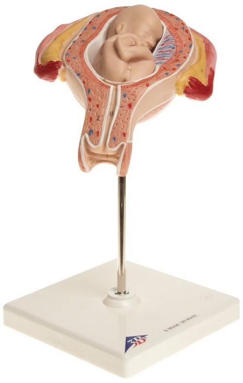
Let’s try to go broke, what is the pelvic presentation of the fetus. This is the location of the fetus in the uterus, in which its pelvic part is located below. It turns out the baby sits with the pelvis down, and not the head, as is necessary for the standard birth process. Such births do not happen very often, in 3-5% of cases. Childbirth in the pelvic presentation of the fetus is considered a pathology, the baby may be injured, complications are possible in the process of labor.
The diagnosis can only be made after 32 weeks of gestation. The baby is in free swimming with her mother in the tummy and turns over as he pleases. Thanks to the intense movement and space in the uterus, the baby develops, its muscles and motor apparatus are strengthened.
How pregnancy proceeds with pelvic presentation of the fetus
Pregnancy with pelvic presentation of the fetus is no different from pregnancy, with head presentation of the fetus. As prevention, special exercises are used. Starting from 32 weeks, you can start a set of exercises developed by professionals and approved by gynecologists. Methods for an obstetric coup have been developed – this is a compulsory way to force a child to adopt the correct birth position. There are circumstances when the children again take the pelvic presentation of the fetus. Hospitalization with pelvic presentation of the fetus is necessary from 38 weeks to diagnose and choose a strategy for conducting labor.
Classification of fetal presentation
Throughout pregnancy, the baby takes various positions, turns over, dances with her mother to music, plays sports and swims. At 32-34 weeks, he begins to actively prepare for childbirth, there is already little space in the uterus, as the baby’s body weight has increased significantly. In most cases, the fetus occupies head diligence, which can be occipital, parietal, facial and frontal. The best option of patrimonial activity – an occipital head presentation. But there is a classification of the location of the fetus in the uterine cavity.
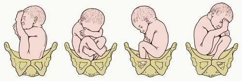
With gluteal presentation of the fetus, the child’s legs are bent at the knees and pressed to the stomach. With the foot – it is the leg is aimed at the exit from the uterus. With mixed presentation, both the knees and hips are bent. In the latter case, they often offer an exercise. In all other cases, you can give birth on your own, but childbirth will be a little more difficult than with the head position of the fetus. Listen to the advice of doctors. Do not be afraid to give birth is not dangerous. All women must go through this.
What are the causes of pelvic presentation of the fetus
Pelvic presentation can be due to the following reasons:
- low water and polyhydramnios of amniotic fluid,
- narrow basin
- the placenta adherence is very low,
- umbilical cord entanglement (double / triple),
- short umbilical cord
- saddle and two-horned uterus,
- uterine defects
- decreased tone of the uterus,
- uterine fibroids,
- fibroma,
- the presence of scars
- multiple pregnancy.
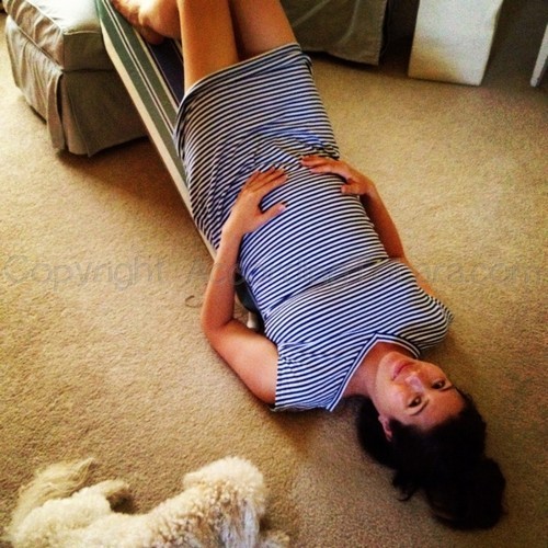
Gymnastics with pelvic presentation of the fetus
Obstetrician-gynecologists have developed special exercises for pelvic presentation of the fetus, with which you can give the baby a head position. You can use such exercises only after the recommendation of the doctor leading your pregnancy.
Exercise number 1
These exercises are performed using flips lying side to side. You need to do 3-5 approaches after a short period of time, after about 8-10 minutes. Do this exercise 3 times a day.
Exercise number 2
Lying on your back, put a cushion or pillow under your lower back. The pelvis should be above the head. This position should be held for 10 to 15 minutes. No longer needed.
How to do the exercises correctly
All exercises should be performed on an empty stomach, so as not to provoke heartburn, nausea, vomiting, severe dizziness. Such exercises are forbidden to be performed earlier than 32 weeks of pregnancy and without consulting a doctor, as well as with late toxicosis, uterine scars, after any gynecological operations. There are also methods of traditional medicine, aromatherapy, which contribute to a change in the position of the fetus in the pelvic presentation and increase the possibility of a head position in the mother’s uterus.
Many fitness clubs and groups for pregnant women offer sets of exercises, including an obstetric coup. You can take this opportunity, but be sure to ask the gynecologist for permission to do such exercises.
Fitness instructors undergo special highly qualified training for working with pregnant women. They clearly know which exercises can be performed and which are prohibited. During the entire period of pregnancy, exercises on the abdominal muscles and oblique muscles are not allowed. Such exercises in the period before conception strengthen the muscles of the abdomen and the press, which greatly improves the process of pregnancy and childbirth.
But Kegel’s exercise will help strengthen the birth canal, make the vaginal muscles more elastic, which will favorably affect the birth process. Do not experiment with your health and the health of your child, do not perform unknown and difficult exercises for you. If you can go swimming and spend more time on the air. Do not forget about breathing exercises.
Ultrasound and medication
If the methods recommended in the article did not achieve the goal, then a pregnant woman can be offered an ultrasound procedure and medical intervention. Usually it is carried out no earlier than 34 weeks of pregnancy with the use of special medications. This procedure is not simple, but it is very effective. After passing it, a woman has the opportunity to give birth in a natural way. This procedure has a number of contraindications: the age of the nulliparous woman is more than 30 years, scars on the uterus, gestosis, entwining the fetus with an umbilical cord, obesity and many other contraindications. This method is not suitable for everyone, therefore it is rarely used in gynecology and obstetrics.
Childbirth with pelvic presentation of the fetus
In the process of labor, the child moves through the birth canal, making a huge number of rotational and translational movements. The baby has to go a difficult way, he gets tired in childbirth no less than his mother, and maybe even more. Therefore, the newborn baby sleeps fast for the first day, sometimes does not even wake up to eat. He needs rest, he has done his first serious work and is now resting calmly and getting ready to meet his parents. Being born is hard work for mom and baby. Life is not easy, and it must be appreciated.
The biomechanism of childbirth with pelvic presentation of the fetus:
- The biomechanism allows you to insert the buttocks into the pelvis of the woman in labor, then the fetus moves along the birth canal. In the process of labor, the buttocks of the fetus fall lower and lower. One of the buttocks is the leading one; subsequently, a birth tumor appears on it, like a fontanel during head insertion.
- Using a biomechanism, the buttocks of the fetus are rotated in the pelvic area, and the gluteal sulcus is created in the right size. One buttock moves to the bosom, the second goes towards the sacrum.
- Cracking of the buttocks after this occurs after fixing the point under the pubic arch, around which the buttock cuts. With a full pelvic position, the buttocks appear first, then the body to the navel, and then the legs. With mixed pelvic diligence with the buttocks, the legs are also born. Handles appear along with the chest, then the shoulders and head.
- The head is born in a bent position. The fixation point is fixed under the pubic arch. Then the chin, face and back of the head appear. The head is born last, the birth tumor does not appear on it.
- The biomechanism of labor during foot presentation is similar to the gluteal. With insufficient opening of the cervix after the amniotic fluid is poured out, the baby’s leg can fall into the vagina, which can slightly delay and complicate the birth process.
Pelvic Prevention
Patients at risk: pelvic presentation, preventive measures should be taken. Pregnant women should follow the regime, have a full 8-hour sleep at night and rest during the day. Particular attention should be paid to nutrition. It should be balanced, consisting of vegetables, fruits, meat, fish, dairy products and cereals. Food should be complete and proper, but do not overeat. A large fetus can complicate labor. It is necessary to carry out prevention to stabilize the nervous system. Spend more time in the fresh air, do what you love. Prevention is also needed to prevent the abnormal activity of uterine contractions. Starting from week 22, the attending physician can prescribe an antispasmodic course for prevention, which can eliminate the causes of pelvic presentation of the fetus.
Maternity clinics offer courses for pregnant women, which detail the rules that should be followed. They carry out psychotherapeutic preparation for childbirth. Training in respiratory gymnastics during childbirth is carried out, recommendations are given for alleviating pain, relieving nervous tension, and controlling the tone of the vaginal muscles.
Pregnancy is a great time for any woman, a new life is growing in her. Any difficulties make us only stronger, and the long-awaited baby becomes the most beloved man in the world. Pelvic presentation of the fetus should not affect your mood. The baby feels all the emotions of the mother and should be happy. Have a good birth. I wish you and your families happiness!