
The kidney is a complex apparatus that performs a number of vital functions that ensure the normal functioning of the body. Each structural element of the body involved in the process of filtration of blood plasma, which formed a waste product – urine.
There are many diseases of the kidneys, chronic or acute which has a damaging effect on a particular structural element, causing diffuse changes of the kidneys.
To understand what is diffuse changes, it is necessary to understand the functional structure of the renal apparatus.
The structure of the kidney
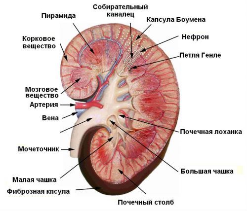
When studying the structure of the kidney, in turn allocate the parenchyma (primary renal tissue) and Cup-pelvis-plating system (CHLS). In the structure of the parenchyma should be allocated to cortex is composed of nephrons (glomeruli surrounded by capsule), and the medulla, consisting of urinary tubules, it is here that the formation of urine. Cup-pelvis-plating system is used for the accumulation and excretion of urine formed.
Arterial blood passes through the long and thin arteries that form the glomerulus, where filtration occurs primary, and then enters the urinary tubules, which provide the absorption (reabsorption) of filtered nutrients (glucose, vitamins, minerals). This ensures maximum cleansing of the plasma and minimizing the loss of nutrients.
Any changes in the structure of the kidney in 90% of cases are a consequence of pathological processes, leading to disruption of their functions. Therefore, the detection of diffuse abnormalities in the tissue during the ultrasound examination, or other diagnostic procedures, is required to perform complex diagnostic procedures aimed at identifying the causes of these changes.
Important! Through the kidneys daily is 150 to 180 liters of fluid that form as a result of reabsorption of 1.5–1.8 liters of urine.
Changes
Detection of diffuse changes cannot be considered a diagnosis, since any structural abnormalities in the tissues of the body are a Testament to the influence of certain pathological processes, which is kidney failure.
Depending on the location of diffuse changes of the following structural abnormalities:
- the body by the kidneys;
- parenchyma;
- sinuses;
- Cup-pelvis-plating system.
Significant role in the diagnosis, plays the character of structural changes, which permits to carry out initial diagnostic verdict, not excluding, however, further comprehensive examination.
For example, you may notice the following deviations:
- change the size of the kidneys;
- the asymmetry of the contours of the kidneys;
- thinning or thickening of the renal parenchyma;
- the formation in the parenchyma of pockets of high or low density;
- abnormalities in the structure of the sinuses;
- changes in the structure of the Cup-pelvis-plating system;
- the liquid in the pyelocaliceal system;
- seals in the structure of the renal vein.
Important! The increase in the Cup-pelvis-plating system in a newborn baby is not a pathology, as occurs as a result of forced accumulation of metabolic products inside its own body, in the period of intrauterine existence.
The lobed structure of the kidney in children is not a pathology, as it bounces to 2-3 years of life
Ultrasound diagnostics
Ultrasound, today, retains a primacy among all the diagnostic procedures in view of availability, high information content, and lack of contraindications for unlimited number research. The method is based on the properties of the soft tissues to resist the penetration of ultrasonic waves.
While some amount of wave is reflected and some passes through the tissue, being absorbed by them. The more reflected ultrasound (hyperechoic area), the lighter the shade on the screen and, consequently, a higher density is on or enable it.
Classification of diffuse changes of kidney structure, with the point of view of the ultrasound examination includes the following types of changes:
- clear;
- fuzzy;
- moderate;
- weak;
- expressed.
Diffuse changes in CHLS, caused by deformity of the pelvis or the renal sinus formed calculi, on the monitor of the ultrasound machine will appear as hyperechogenic areas. Tissue having a low density, ULTRASONIC monitor will affect how the darker areas are called hypoechoic. The fluid contained in the structure body, for example, the cyst, is characterized as anahowana education.
Diffuse changes in the kidneys with ultrasound will have the following characteristics:
- the shadows in the parenchyma;
- hyperechogenic areas in CHLS;
- the lack of clear contours in the parenchyma;
- the shadows in the contours of the renal arteries
- anahowana areas in the parenchyma or CHLS;
- deformation of the contours of the pelvis and the renal capsule.
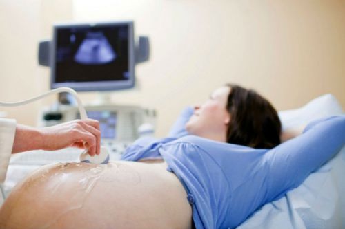
Using ultrasound to explore the functional state of the kidneys in the fetus in late pregnancy
Reasons
The causes of deterioration of kidney structure can lie in a variety of pathologies or anatomical changes in congenital or acquired. For example, a congenital bend of the ureter or curvature as a result of squeezing it in the process of pregnancy, the growing fetus may lead to development of hydronephrosis.
The increase in the Cup-pelvis-plating system, which is a direct symptom of the disease at diagnosis, defined as “diffuse changes of CHLS”.

Structural changes CHLS and sinuses kidneys can cause:
- cystic masses in the cavity of the pelvis or cups;
- stones in CHLS;
- a nodule.
The defeat of the sinus of the kidney is often accompanied by pain in the heart and increased blood pressure
Of great importance in the development of structural damage of the sinus of the kidneys are inflammatory and sclerotic processes that cause the swelling (in the case of inflammation) or atrophy (in the case of atherosclerotic changes) in the vascular surface of the sinus. Often, inadequate treatment of inflammatory diseases leads to the development of atrophic processes.
Important! A characteristic feature accompanying the course of acute stage of disease is the increased authority, while the chronic – on the contrary, its decreasing.
Diffuse changes of renal parenchyma may have different manifestations in connection with the structural feature of parenchymal tissue.
In the list of diseases that cause disruption of the normal structure of renal tissue, included:
- parenchymal cyst;
- pyelonephritis;
- glomerulonephritis;
- nephrosclerosis;
- TB.
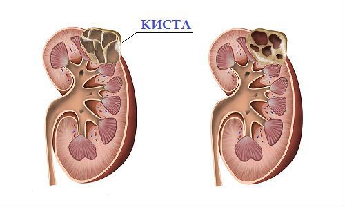
Parenchymal cyst
Parenchymal cyst of kidney is a congenital or acquired pathology, in which, directly in the main fabric body formed a cavity space filled with a serous or hemorrhagic exudate. A cyst can form in the kidney in a single instance (solitary), but may be multiple cystic lesions (cystic).
Great diagnostic importance is the mechanism of formation of cysts. If cystic cavity formed as a result of injury or violation of the outflow of fluid from the channel of the nephron obstruction due to uric acid crystals, as a rule, such education is benign and easily removed laparoscopiceski method. In the diagnosis of a cyst is defined as a single cavity, round or oval with clear boundaries, filled with liquid contents.
The overlap of the lumen of the canaliculus polyps or dysplastic changes, such as proliferation of connective tissue leads to the formation of multilocular cysts, which is a multi-cavitary education with clear contours.
The multi-cell cyst is considered to be the cystic form of cancer
Pyelonephritis
Inflammatory disease of the kidney associated with damage to the renal tissue and the renal pelvis. Most often the disease affects both right and left kidneys and can be acute or chronic.
The reasons for the development of pyelonephritis may serve as:
- Adenoma of the prostate gland. Enlarged gland impedes the timely flow of urine from the kidneys, contributing to the development of the inflammatory process. Also playing a role, kidney infection of pathogenic microorganisms, developing in the prostate gland;
- Vesico-ureteral reflux. In most cases, the development of pyelonephritis is preceded by cystitis or urethritis. As a result of prolonged course of these diseases disrupted the mechanism that prevents back urine reflux into the ureter, which is the cause of infection of the kidneys;
- Urolithiasis. In addition to violations of the outflow of urine, stones damage the mucosa of the pelvis, contribute to the penetration of pathogens into the tissues of the kidneys.
When visualization of the kidneys on the ULTRASOUND monitor, in acute course of the disease, an increase in the thickness of the parenchyma, the disparity in size of both kidneys, and for chronic uneven contours, heterogeneity (due to scarring) and thinning of the structure of the underlying tissue. In chronic pyelonephritis observed an extensive diffuse changes in the parenchyma.
Glomerulonephritis
Usually glomerulonephritis occurs as a result of previously transferred infectious diseases:
- sore throat;
- otitis;
- scarlet fever;
- pneumonia.
The immune changes in the body, triggered by bacterial microflora, forces us to accept its own tissues of the kidneys as an alien, exposing them to the attack of the protective systems. In a healthy organism, the immune complexes must be neutralized in the liver, if this does not happen, the vessels of the glomeruli exposed to the ravages.

In glomerulonephritis, the kidney size is usually normal, but may be increased. The structure of the parenchyma is uneven, due to the increase of the glomeruli, vascular system is ill defined, there may be multiple microscopic hemorrhages and pericardial cavity.
Hematuria is one of the diagnostic signs of glomerulonephritis
Nephrosclerosis
Nephrosclerosis is a disease associated with insufficient blood supply to the kidneys due to the sclerotic lesions of the vascular system. Violation of blood supply leads to a gradual withering away of the functional components of the kidney – the glomeruli and a gradual replacement of connective tissue.
Due to the fact that the interstitial tissue has a dense structure, the intensity of the ultrasound has some differences, making are defined by extensive diffuse changes. In addition, when nephrosclerosis occur atrophic changes that reduce the size of the body (shrinkage) and the thinning of its sheath.
Stage nephrosclerosis is divided into:
- primary;
- secondary.
Atherosclerotic vascular changes are a major cause of development of primary nephrosclerosis
If the conclusion of the medical examination, there is the wording “primary wrinkled kidney” – it means the pathological processes caused by atherosclerotic lesions of the vascular system.
Secondary wrinkled kidney is the result of chronic inflammation with irreversible parenchymal damage:
- glomerulonephritis;
- tuberculosis;
- pyelonephritis.
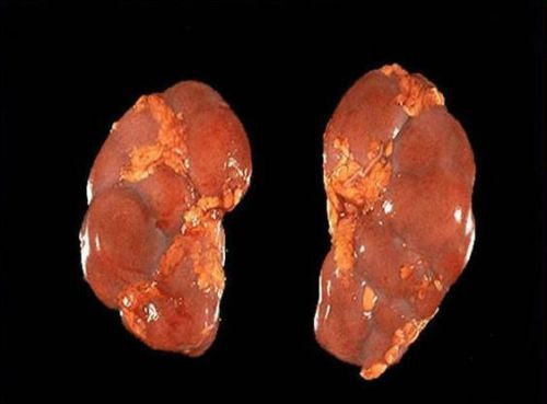
Important! When the nephrosclerosis, kidneys are fine-grained surface, resulting in ultrasound contour is indistinct, in some cases, determined by a pronounced tuberosity.
Tuberculosis
Tuberculous lesions of the kidney, depending on the stage of development can have various manifestations:
- multifocal lesion total volume of the parenchyma, accompanied by the formation of capsules, filled with necrotic masses. Ultrasound capsules are defined as multiple cystic masses filled, unlike cysts, not exudate, and more dense masses (caseous);
- isolated isolated foci of parenchymal damage;
- multiple scarring (areas of increased echogenicity). This process occurs when you restore the body after the disease;
- partial or complete substitution of one of the segments of healthy tissue kidney encapsulated foci of necrosis;
- lose more than 70% of the body tissues.
Important! In addition to the formed capsules, ultrasound determines the deformation of the Cup-pelvis-plating system, which has certain similarities with similar deformation with the development of parenchymal cysts.
TB treatment requires a complex approach, due to the rapid development of resistance of the Bacillus to applied preparations.
Thus, the concept of diffuse changes means quite a wide range of structural transformations that have a negative impact on the functional activity of the organ. The main goal of diagnostic procedures is the accurate description of these changes, allowing a higher percentage of accuracy to identify the disease and develop the most effective tactics of treatment.


Hi there The bookmaker is simply a middle-man who operates on a small revenue margin and ideally likes to see a fair overround, assuring a revenue. danke