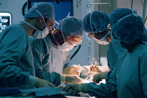
Tumors of the heart are treated surgically. Surgeries for heart tumors can include removal of an intracavitary tumor, removal of a myocardial or pericardial tumor, pericardectomy, removal of a pericardial cyst.
During the radical operation, excision of the heart tumor with the surrounding surrounding tissues, suturing or plastic surgery of the defect are performed. Benign heart tumors in most cases can be removed radically. In some cases, patients may need plastics or valve replacement.
Removal of primary malignant heart tumors is ineffective. In this case, most often resort to partial (palliative) removal of the tumor, followed by radiotherapy or chemotherapy. For secondary tumors of the heart, the treatment is also palliative.
Tumors of the heart – a heterogeneous group of tumors that grows from the tissues and membranes of the heart. Tumors can develop from any tissue of the heart and occur at any age. Neoplasms can sprout heart muscle, pericardium, affect the valves and septum of the heart. In the fetus, they can be detected using fetal echocardiography, starting from 16-20 weeks. intrauterine development. Primary cardiac tumors are found in cardiology with a frequency of 0.001-0.2%; secondary (metastatic) – 25-30 times more often. All tumors of the heart carry the potential danger of mortally dangerous complications – heart failure, arrhythmias, pericarditis, cardiac tamponade, systemic emboli.
Tumors of the heart – heterogeneous on the histological structure of the tumor, primarily arising from the tissues of the heart or germinating in them from other organs. Tumors of the heart, depending on their type, location and size, can cause shortness of breath, cough, tachycardia, arrhythmias, chest pain, heart failure, cardiac tamponade, thromboembolism. Diagnosis of heart tumors is carried out taking into account the data of EchoCG, X-ray, ventriculography, MRI and MSCT of the heart, ECG, biopsy. At revealing of primary benign tumors of the heart, their radical excision is performed; treatment of primary malignant and metastatic tumors is usually palliative (radiation therapy, chemotherapy).
Classification of heart tumors
Tumors of the heart, representing independent diseases, are primary; tumors that metastasize through the blood and lymphatic vessels or germinate from neighboring organs are secondary. The causes of the development of primary heart tumors are unknown. Secondary tumors of the heart are more often represented by metastasis of breast, stomach, and lung cancers, and more rarely thyroid and kidney cancers.
According to the morphological principle, heart tumors are divided into benign (make up 75%) and malignant (make up 25%). By origin, malignant neoplasms can be both primary and metastatic, secondary.
Among benign tumors are myxoma of the heart (50-80%), teratomas, rhabdomyomas, fibromas, hemangiomas, lipomas, papillary fibroelastomas, pericardial cysts, paragangliomas, etc. Malignant neoplasms include sarcomas, pericardial mesothelioma and lymphomas.
Pseudo-tumors include foreign bodies of the heart, organized blood clots, inflammatory formations (abscesses, gummas, granulomas), echinococcal and other parasitic cysts, and calcification conglomerates. A separate group consists of extracardiac tumors of the mediastinum and pericardium, which squeeze the heart.
Primary benign heart tumors
Myxomas
Half of all primary heart tumors occur in myxoma. Sporadic myxoma of the heart is 2-4 times more often found in women.
There is a hereditary Karni complex with an autosomal dominant type of inheritance, characterized by multicentric tumors of various localities – myxoma of the heart, pigmented skin tumors, breast fibroadenomas, ovarian cysts, nodular dysplasia of the adrenal glands, myxoid testicular tumors, hypophysis adenomas, hysteromyiasis, hypophysis, hysteromyiasis, hysteromyoma
The predominant localization of the mix is the left atrium (about 75%). Myxomas with a leg prolapse through the mitral valve, making it difficult to empty the left atrium and fill the ventricle during diastole. Macroscopically, myxomas can have a slimy, hard, lobed or friable structure. Unformed loose myxoma represent the greatest danger in terms of the development of systemic embolism.
Papillary fibroelastomas
Among the primary tumors of the heart, benign papillary fibroelastomas, mainly affecting the aortic and mitral valves, are second in frequency.
Morphologically, they represent avascular papillomas with branches resembling anemones extending from the central nucleus. Most often they have a leg, but unlike the mix, they do not cause valve dysfunction, but they increase the likelihood of embolism.
Rhabdomyoma
Rhabdomyomas among all benign heart tumors account for 20% and are the most frequent neoplasms in children. Usually, rhabdomyomas are multiple, have intramural localization in the septum or wall of the left ventricle, and affect the cardiac conduction system.
The course of rhabdomyomas may be accompanied by tachycardia, arrhythmias, and heart failure. These heart tumors are often associated with tuberous sclerosis, sebaceous gland adenomas, benign renal neoplasms.
Fibroids
Connective tissue tumors of the heart are also mainly found in children. They can affect the valves and the conducting system of the heart, cause mechanical obstruction, imitating valvular stenosis, the clinical picture of heart failure, hypertrophic cardiomyopathy, constrictive pericarditis.
Heart fibroids can be part of the syndrome of basal cell nevus (Gorlin syndrome).
Other benign heart tumors
Hemangiomas are found in 5-10% of cases of all primary heart tumors. More often they do not cause clinical symptoms and are detected during a routine examination. Less commonly, intramyocardial hemangiomas are accompanied by impaired atrioventricular conduction, and during germination of the atrioventricular node may lead to sudden death.
Lipomas of the heart can develop at any age. Usually these are tumors on a broad basis, localized in the epicardium or endocardium. The course of lipomas is often asymptomatic; upon reaching a large size, they can cause arrhythmias, conduction disturbances, a change in the shape of the heart, radiologically detectable.
Pheochromocytomas may have intrapericardial or myocardial localization, accompanied by the secretion of catecholamines. Pericardial cysts on chest radiographs often mimic heart tumors or exudative pericarditis. Most often they are asymptomatic, sometimes they can cause symptoms of compression of the chest organs.
Malignant heart tumors
Sarcomas are the most common primary malignant heart tumors. They occur mainly at a young age (average age 40 years). Heart sarcomas can be represented by angiosarcomas (40%), undifferentiated sarcomas (25%), malignant fibrous histiocytomas (11-24%), leiomyosarcomas (8-9%), rhabdomyosarcomas, fibrosarcomas, liposarcomas, osteosarcomas. Malignant tumors of the heart often occur in the left atrium, leading to obstruction of the mitral orifice, cardiac tamponade, heart failure, and lung metastasis.
Pericardial mesothelioma is relatively rare and occurs mostly in men. They usually metastasize to the pleura, spine, brain, surrounding soft tissue.
Primary lymphomas most commonly affect immunocompromised individuals (including HIV infection). These tumors of the heart are prone to extremely rapid growth and are accompanied by heart failure, arrhythmias, tamponade and superior vena cava syndrome.
Metastatic tumors of the heart most often affect the pericardium, at least – the heart muscle, endocardium and heart valves. Like primary heart tumors, they can cause shortness of breath, acute pericarditis, cardiac tamponade, rhythm disturbances, atrioventricular block, and congestive heart failure. In the heart, lung cancer, breast cancer, renal cancer, soft tissue sarcoma, leukemia, melanoma, lymphoma, Kaposi’s sarcoma can metastasize.
Symptoms of heart tumors
Manifestations of tumors of the heart due to the type of neoplasm, its localization, size, ability to decay. Extracardiac tumors are manifested by fever, chills, weight loss, arthralgia, skin rashes. When a tumor is compressed by chambers of the heart or coronary arteries, shortness of breath and chest pain occur. Tumor growth or bleeding can cause cardiac tamponade.
Heart tumors with intramyocardial growth (rhabdomyomas, fibromas) that are squeezed or inserted into the conduction system are accompanied by atrioventricular or intraventricular block, paroxysmal tachycardias (supraventricular or ventricular).
Intracavitary tumors of the heart mainly disrupt the function of the valves and impede the flow of blood from the chambers of the heart. They can cause the phenomenon of mitral and tricuspid stenosis or failure, heart failure. Symptoms of intracavitary tumors of the heart usually occur when the position of the body changes due to changes in hemodynamics and physical forces acting on the tumor.
Often the first manifestations of heart tumors are thromboembolism in the vessels of the systemic or pulmonary circulation. Tumors from the right heart can cause pulmonary embolism, pulmonary hypertension, and pulmonary heart; tumors of the left heart – transient cerebral ischemia and stroke, myocardial infarction, limb ischemia, etc. The occurrence of infarctions of internal organs in young people in the absence of congenital and acquired heart defects, atrial fibrillation and infective endocarditis makes you think about the presence of a heart tumor.
Diagnosis of heart tumors
Due to the variability of the clinical picture and the multiplicity of the morphological forms of heart tumors, their diagnosis is not an easy task.
ECG data for heart tumors are polymorphic and low-specific: they can reflect signs of hypertrophy of the heart chambers, conduction and rhythm disturbances, myocardial ischemia, etc. Chest X-rays often reveal an increase in heart size and signs of pulmonary hypertension. A more sensitive method for the diagnosis of tumors is an ultrasound of the heart: atrial transshoracic echoCG, a ventricular tumor, is visualized by transesophageal echoCG.
In case of doubtful diagnostic results, MRI and MSCT of the heart, radioisotope scanning, heart cavity sounding and ventriculography are performed. To verify the histological structure of the heart tumor, a biopsy is performed during catheterization or diagnostic thoracotomy.
In exudative pericarditis, cytological examination of the fluid obtained by pericardial puncture can provide valuable diagnostic information. Differential diagnosis of heart tumors is carried out with CHD, myocarditis, cardiomyopathy, pericarditis, amyloidosis of the heart.
Prognosis of heart tumors
Removal of single benign heart tumors is usually accompanied by good separated results – 3-year survival is 95%.
In the future, patients are shown to observe a cardiologist and a cardiac surgeon with an annual echocardiography to detect the recurrence of a heart tumor in time. Multiple heart tumors reduce survival over the next 5 years to 15%.
In primary and metastatic tumors of the heart, the prognosis is very poor; surgical treatment is ineffective, chemotherapy and radiation therapy have little effect on the prognosis.