The question of treatment of osteoarthritis of the knee has become a byword. The problem is very common. And, oddly enough, is ambiguous.
The ambiguity lies in the approaches to treatment.
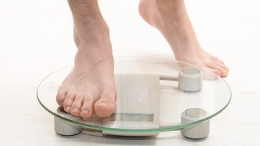
What are the challenges when newly diagnosed osteoarthritis? First, we should examine whether osteoarthritis is a cause of concern for the patient?
Often the situation occurs in the following scenario:
- Patient feels pain in the joints (in the absence of injury), he turns to the doctor.
- The doctor prescribes examination (usually x-ray, ultrasound sometimes additional).
- The x-rays show signs of osteoarthritis.
- Treatment.
The result is often appear on the admission of patients is quite young (25-35 years) with the question on how to treat arthritis as assigned earlier the treatment gives only a temporary effect. The weak link in this scheme is a method of diagnosis.
Even at the joint, which revealed only the initial signs of osteoarthritis on x-rays in addition to add anything more in the conclusion of the radiologist mentioned will not. It is not surprising: in addition to traumatic injuries and signs of osteoarthritis on an x-ray is rare to see any changes. By ultrasound in most cases we can reveal inflammatory changes, but this is not always enough to understand the situation.
The fact that for young patients (under 35 years) are the most common causes of joint pain not osteoarthritis. The knee joint has a fairly complicated structure, with lots of ligaments, menisci, and shells. And to identify, for example, damage to the meniscus of the knee joint, which happens quite often, during the examination of the x-ray and ultrasound impossible. The fact is that on the ultrasound the meniscus is only partially visible, and x-rays are not visible at all.

What research is optimal in this case? Best MRI. This is the kind of research where we can evaluate not only the condition of the bone structure and soft tissues. However, it is worth noting: the menisci are small enough and resolution low-power devices (less than 1 Tesla) is clearly insufficient to obtain the desired information.
When using MRI we can identify a number of problems such as:
- damage to the meniscus;
- ligament damage;
- inflammatory changes;
- fragments in the joint cavity, etc.
Why not always, the patient receives direction on this study? The reasons are usually two: either the well-established legacy scheme of examination and treatment used by the attending physician, or forced circumstances (no MRI in the institution, a limited number of coupons for directions, etc.).
In addition, the source of pain is not always the joint itself, so we cannot say that in each case the presence of pain in the joint you need to run an MRI. Often the source of pain may be the muscles and their tendons around the joint. Such phenomena are usually seen during the inspection and run some tests.
Many of these issues can really be detected at the initial examination. This may help suggest a particular diagnosis, and to choose the right survey method, which helps to decrease the time until diagnosis and appropriate treatment.