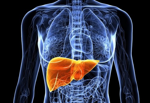
Hemangioma of the liver – this mysterious tumor, reasons for the emergence and growth which still remain a mystery for physicians. Hemangioma affects approximately 2% of the population, and in men it occurs less frequently than in women: one man with hemangioma have 4-6 women.
To be a hemangioma of the liver at any age, but in most cases it is recorded among persons 30 to 50 years. In women the age limit is shifted to the bottom – they have more common hemangioma in a young age, and the tumor is usually of larger size.
Liver hemangiomas can be found in infants, in some cases, they even were prenatally detected in a growing fetus! Fortunately, hemangioma liver always benign; cases of transformation into malignant tumor were noted.
What is a hemangioma of the liver and its causes
Strictly speaking, a hemangioma is a cluster of vessels, which is endotelialny tapered tube divided by fibrous septae. Usually liver hemangioma only one; cases where there are several, are quite rare. The size of hemangiomas can range from small masses on liver size of 2 mm to giants of more than 20 cm At a superficial inspection of the surface of the hemangioma of the liver can be both flat and bumpy due to formation damage (hematoma).
One of the main signs hemangioma – color; it it reddish-blue and well distinguishable from the surrounding liver tissue. Large tumors can be on the leg. The right lobe of the liver more susceptible to the formation of hemangiomas than the left.
Hemangioma of the liver can be as cavernous (several large cavities with inner walls of fibrous tissue), and in much more rare cases, capillary (many small cavities, each of which contains the vessel). The last diagnose is very difficult – they are so small that are not visible during the examination.
Causes of liver hemangiomas remain a mystery for doctors, although it is assumed that hormonal contraceptives and steroids may accelerate its growth (it is unclear, however, whether these drugs contribute to her education). It is also unknown whether there is genetic predisposition to the appearance of hemangioma of the liver; there is evidence, however, rare that women of different generations in one family had the hemangioma. Some scientists believe that liver hemangioma is a benign congenital hamartoma (tissue abnormality). Hemangiomas can be laid in childhood, and occur under the influence of yet unknown factors.
More often occur hepatic hemangioma of skin; however, it is unclear, do they influence in any way on the existence of their “sisters” in the liver.
Symptoms of hemangioma liver
Usually people with liver hemangioma don’t feel any discomfort and especially not require treatment. The presence of hemangiomas are most often discovered by chance during medical examinations in cases of suspected other disease, or after death at autopsy. However, after the diagnosis of hemangioma of the possible discomfort of the psychological plan – a person can be frustrating at the thought that his liver is tumor. However, you should not be afraid, hemangioma of the liver – the tumor is benign and rarely cause inconvenience to its owner.
If symptoms still are present, patients often complain of pain in the right upper abdomen. In some cases the occurrence of pain is explained by thrombosis, krovoisliania due to rupture or mechanical compression of neighboring vascular hemangioma liver organs. In other cases the cause of pain cannot be established. Sometimes the liver of the patient increases, however, when a hemangioma is a rare occurrence. Patients may complain of a feeling of fullness after eating small amounts of food, nausea, vomiting. As a rule, the larger the hemangioma of the liver, the more pronounced the symptoms – 40% of people with hemangioma is 4 cm, up to 90% of people with hemangioma the size of 10 cm.
However, these signs are not specific for hemangiomas of the liver and can be caused by other diseases. In any case, a constant feeling of discomfort and abdominal pain should see a doctor.
The factors causing the growth of hemangioma liver
According to existing research, can affect the growth of hemangiomas may be the steroids and the stimulation of yashnyky horionichesky gonadotropin human. Women who took hormone replacement therapy, hemangioma of the liver were found more often. Another risk factor is pregnancy – pregnant hemangiomas are detected more frequently than never beremennyh. It is believed that role plays estrogen, that a woman’s body increases during pregnancy.
In 40% of cases of hemangioma of the liver increase in size with a small speed – up to 2 mm per year. Faster the tumor is growing young, not older than 30 years, and slowest – the people who turned the half-century milestone. The more hemangioma of the liver, the slower it grows.
Hemangioma of the liver in children
One of the most common types of tumours in infancy is a hemangioma. Approximately 5-10% of children under the age of one year are observed hemangiomas, which are then in most cases (80%) required no treatment and was held safely by themselves. More often, however, hemangiomas appear on the skin and subcutaneous tissues, but sometimes affect the liver.
Hemangioma of the liver in pregnant women
If a woman was diagnosed with a hemangioma and then she got pregnant, the risk of complications related to the neoplasm, increases. As expected, the increased amount of estrogen causes an increase in liver hemangiomas.
In rare cases, growing hemangioma of the liver may require treatment. A woman may observe such signs as pain in the upper right abdominal region, bloating, nausea. However, the presence of the hemangioma does not mean that a woman cannot get pregnant; it is best to discuss this with your doctor.
Drugs that affect hormone levels in the body (for example, birth control pills) can cause complications when diagnosed with hemangioma of the liver. Again, this question is best discussed with your doctor.
Complications of hemangioma liver
In General, the prognosis in a person with hepatic hemangioma is very good. Not yet met cases when the hemangioma of the liver would be reborn into a malignant cancer. However, in some cases, possible complications, largely depending on the size and location of the tumor. It is worth noting that such cases are very rare.
Rupture of the tumor. In rare cases of large cavernous hemangioma of the liver can rupture by themselves or as a result of injury, which can lead to vascular shock, or hemoperitoneum (when the blood flows into the peritoneal cavity). The man turns pale, his disturbed heart rhythm, decreases blood pressure, the skin performs a cold sweat. If you notice such symptoms should immediately seek emergency medical care is a condition threatening the patient’s life and requires immediate treatment.
Intratumoral bleeding. It can be diagnosed by the presence of feces in the blood trapped there as a result of falling from the liver into the intestine via the bile ducts.
Compression of the bile duct near the tumor arteries and veins. In rare cases, the swelling can pinch the nearby blood vessel, causing the blood circulation (for example, when overlapping a hepatic hemangioma of the inferior Vena cava there is swelling of the legs).
Compression of the stomach. In very rare cases, a hemangioma of the liver may compress the stomach, preventing the exit of its contents. At the same time develop the following symptoms: rapid saturation when taking even small amounts of food, nausea, vomiting, stomach discomfort
Hemobilia. Another rare disease in which the blood through the bile ducts gets into the intestine. In humans, there may be yellowness of the skin and mucous membranes, dark urine, discoloration of feces.
In rare cases, multiple hemangiomas of the liver is large in size may require surgical treatment.
Diagnosed with hemangioma of the liver
Hemangioma is one big diagnostic problem. Diagnosis is complicated by the fact that near the hemangioma can be (and sometimes imitating it) other liver lesions, both benign and malignant. Therefore, it is necessary to differentiate a hemangioma from a benign (cysts, adenomas, focal hyperplasia nodes, abscess) and malignant tumors (carcinoma, hepatic angiosarcoma, metastases in the liver).
In some cases of hemangioma of the liver – a consequence of the development of other diseases, for example, when Klippel-Trenaunay-Weber syndrome or Kasabach-Merritt. It was reported multiple hemangiomas of the liver in patients with systemic lupus erythematosus.
Examination hemangioma of the liver
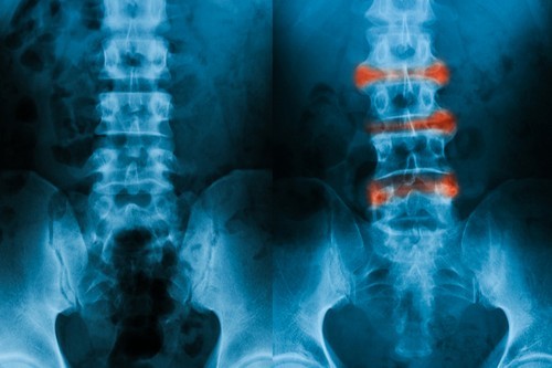
On physical examination to probe the hepatic hemangioma is impossible due to its small size. Blood, urine and feces when hemangioma liver completely normal (although in rare cases, large tumors may present with thrombocytopenia). Therefore, for the diagnosis of several commonly used laboratory and instrumental methods, such as ultrasound, computed tomography, magnetic resonance tomography and hepatic arteriography.
Ultrasound. Widely available, cheap and harmless method of examination. Liver hemangiomas are usually echogenic, but the best results are obtained with color Doppler mapping. If the performance of conventional ultrasound for the determination of liver hemangiomas is 46%, in the DRC, it increases to 69%.
CT. For examination of hemangiomas, it is preferable to study with a contrast agent; a hemangioma of the liver could correctly identify 66% of cases.
Magnetic resonance imaging. As with CT, the best liver hemangiomas are defined when entering into the patient a contrast agent. However, small hemangioma (less than 2 cm in diameter) in the picture look like carcinoma or liver metastases, but overall the performance testing are very high – above 90%. However, conventional MRI of benign hemangioma of the liver may be much more dangerous hepatic angiosarcoma. One of the main differences from a traditional liver hemangiomas is rapid growth, so your doctor may suggest the patient to undergo MRI again in a few months to determine the rate of tumor growth.
Single photon emission computed tomography (SPECT or SPECT) is a relatively recent invention. Unlike previous surveys, this technique allows you to create three-dimensional images. It can help to diagnose the hemangioma of the liver more efficiently; unfortunately, emission tomography is not available everywhere. When the diagnosis of hemangioma liver size up to 2 cm sect gives the best results.
Arteriography. Although diagnostic accuracy described above is a non-invasive surveys is quite high, arteriography may be useful in the diagnosis of some hemangiomas. Determine their availability on the displacement of the branches of the hepatic artery, stretching and the degree of fullness.
Biopsy. This method is not recommended because of the increased risk of bleeding when taking material. If it is unclear, hemangioma or cancer, a biopsy is recommended instead to use a combination of computed tomography and magnetic resonance imaging. And only if the survey is unsatisfactory, you can resort to biopsy.
Treatment hemangioma liver
The vast majority of hemangiomas of the liver to the small size, does not cause unpleasant symptoms do not require treatment and those that remain throughout the life of the patient. Studies have shown that only one of 47 patients in rescan in a few years hemangioma liver increased in size. In addition, there are no identified cases where the hemangioma would have degenerated into a malignant tumor. Therefore, doctors usually suggest to undergo a repeat examination in 6-12 months after detection of a hemangioma of the liver, to see if increased it in size. If it does not, as a rule, the doctor recommends to leave it as is. There is no need to treat, and even in further surveys, there is no need.
With the exception of patients who suffer from diseases of the liver, undergo hormonal treatment or become pregnant. Also the condition of patients, liver hemangiomas exceeding 10 cm, you should watch more carefully; usually, they are advised to check the status of your liver every year due to the potential risk of complications.
Drug therapy for hemangioma liver
Until recently, no method existed whereby it would be possible to reduce the size of the hemangioma liver drugs. And only a few years ago, the doctors found that hemangiomas can be reduced with the help of sorafenib (an antitumor drug, the inhibitor multikinase). Now preparations on the basis of sorafenib are widely used in the treatment of renal cell and hepatocellular carcinoma.
Surgical treatment of hemangioma of the liver
Typically, the surgery is prescribed if the hemangioma causes adverse symptoms. Unfortunately, it is difficult to determine who is guilty of a hemangioma of the liver or any other disease (e.g. irritable bowel syndrome). It happens that after the operation, the person continues to feel pain in the abdomen; hence, the reason it was not a hemangioma of the liver, and something else.
Also surgical treatment is indicated if the hemangioma is growing rapidly or if it is impossible to differentiate it from malignant tumors of the liver. And, of course, if ruptured hemangioma – cavernous hemangioma liver rupture heavy bleeding that can even lead to death of the patient. Fortunately, this happens rarely – the risk of rupture of a large hemangioma of the liver is only 3.2%.
The first thing the patient with a ruptured hepatic hemangioma stop the bleeding by ligation or embolization of hepatic arteries. Once the patient is stabilized, surgery is performed to remove the hemangioma. Preference is given to minimally invasive techniques, for example, arterial embolization and sclerotherapy (so effectively just to get rid of many small hemangiomas; although they do not disappear, but their further growth is excluded). Stop the growth of hemangiomas of the liver also contributes to radiofrequency destruction of the.
Doctors can not agree whether to preemptively remove a large hemangioma of the liver. On the one hand, the rupture of the hemangioma threatens the patient’s life. On the other hand, rupture of liver hemangiomas can sometimes be no more than 3.2% of cases, but the percentage of complications after surgery to remove the hemangioma is higher more than twice. After surgery in 7% of patients developed complications, including life-threatening. As a result, doctors tend to think that surgical treatment should be prescribed only to patients with severe symptoms or serious complications of the disease.
Life with hemangioma of liver
People diagnosed with uncomplicated hemangioma liver do not require a special diet, for them there are no restrictions in physical activity they can continue the lifestyle to which you are accustomed, without the slightest damage to itself. However, if the hemangioma is large, the doctor will advise the patient to avoid activities that contribute to the injury of the right upper square of the abdomen, where the liver.