Traditionally used diagnostic methods, such as survey and examination of the patient, auscultation, percussion, palpation, and the use of laboratory methods in the majority of cases insufficient.
To accurately determine the form of disease, and to visually ascertain the pathological changes in internal organs, for many years used different instrumental methods. Of more modern it can be noted computer and magnetic resonance tomography, and ultrasonic scanning.
In addition to all the methods of diagnosis of diseases of the urinary organs is very informative and reliable method is excretory urography.
This method, used since 1929 and is constantly improving, mainly due to the use of the latest solutions for contrast and modern models of x-ray installations, and today remains very popular in urological and nephrological medicine industries.
Data obtained during examination of the patient, require confirmation by instrumental methods
Based on what information in the study
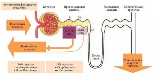
In these two words: “excretory” and “urography” is the essence of the method. Excretory – means “select some stuff”, and urography is that the allocation happens through the kidneys, and whole process is recorded separate pictures (graphy), or sequentially and dynamically (scopy) through x-ray machine. This is possible only when a substance filtered by the kidneys and then coming in the urinary channels, is the contrast, that is able to picture clearly stand out amid all the other internal organs.

His movement through the blood vessels after intravenous injection is very fast, and within 5-10 minutes the contrast enters the renal artery and then the kidney. Being filtrated in the renal glomeruli, a contrast agent becomes an integral component of the urine begins to move down the ureters, fills the bladder and then excreted through the urethra during urination.
During this process, certain moments of which are captured radiographic apparatus at 5-7 minutes 12-15 minutes 20-25, and 46 and 60 minutes according to the testimony, the contrast allows you to define various pathological conditions of the urinary organs: anatomical defects or functional character. Therefore, excretory urography is indicated for a wide range of complaints of the patient and is quite common in children and adults for confirmation or exclusion of various diseases of the kidneys, ureters, bladder, urethra.
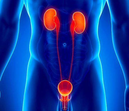
If excretory urography is evaluated all the urinary organs
In addition to receiving this data, revealed by x-rays signs will help professionals to learn about the status of the neighboring kidneys and bladder organs. This is especially important if there is suspicion of a doctor to their negative influence on the urinary system.
That information turned out to be the most reliable and useful for the diagnosis, is very important to choose the contrast agent, which would not have a negative impact on the body of the subject.
What a contrast agent used in renal pathology
These drugs, also known as RKS (contrast media) are used for radiation, that is, conducted using x-ray diagnostics. The value of such information compared to conventional radiography, increases several times. Now developed contrast agents for MRI and ultrasound, but excretory urography of the kidneys, using contrast is a lot more of these methods.
Especially popular rentgenopozitivnye RKS non-radioisotope, which are inhibited by ionizing radiation, making the photographs look clear and white formulations. This is because the density of the RCC is much more bone structures and especially the soft tissues. The more the atomic mass of an element included in the contrast, the more rentgenopozitivee and more readable in the images becomes biological environment of the body that contains it.
Urografin is one of the commonly used radiopaque compounds
Of the chemical elements, the most optimal for human studies of the kidneys was used iodine – or rather its salts, are well soluble in water. But such RKS, called ion, cause a number of side effects: nausea, vomiting, cardiovascular disorders, lowering blood pressure. Therefore, the stakes of nonionic RCM, having low toxicity and excellent wrapping patients. But due to their high value now in conducting excretory urography participate and ionic contrast agents, gives side effects in 12% of cases, and nonionic RCM.
It is possible to mention the following medications:
- ion – Yodamid, Diatrizoate, Iothalamate, Ioxaglate, Iodipamide;
- nonionic – Iopamidol, Iopromid, Iohexol, Iotrolan, Urografin, Iopromid (Ultravist), Iodixanol (Visipak).
These funds differ in that once in the blood stream of a human, they do not bind with plasma proteins and virtually unchanged on the chemical composition of excreted by the kidneys. Despite the availability of modern nonionic contrast agents remains urgent and a security issue in the implementation of the review of excretory urography and its other varieties, intravenous urography (drip introduction of contrast), so in a clinic or hospital should always be provided for measures to provide patient emergency medical care.
The same contrast agents are used for another study of the urinary tract, retrograde urography. Its essence lies in the introduction of contrast through the urethra into the bladder through a catheter. This chemical substance is not absorbed into the bloodstream, that is an advantage of the method, however, the specialist fails to obtain sufficient information about the condition of the kidneys.
In some cases, it is necessary to conduct excretory urography
Details to investigate the excretory function of the kidneys, their anatomy, the structure of the ureters and bladder is necessary in many cases. This study can be assigned as in the subjective complaints of the patient with pain in the lumbar region, and the detection of pathological changes in the urine or in cases of injury.
In General, all indications for excretory urography can be represented as follows:
- pain of any intensity and character, localized in the lumbar and inguinal region;
- pain in the projection of the bladder (suprapubic);
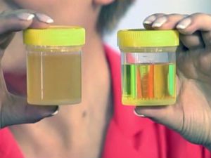
- detection of hematuria, significant (pink or red urine) or weak (some erythrocytes in the urine);
- dysuric disorders: frequency and abundance of urination, soreness or discontinuous;
- recurrent infectious diseases of the urinary channels;
- data for pyelonephritis, urolithiasis or obstruction of the ureters;
- the appearance of local or General edema;
- high blood pressure;
- postoperative complications;
- injuries of the kidneys or bladder;
- obtained with ultrasound of the assumptions about the presence of anatomical defects in the structure of the kidneys and urinary tract.
The results obtained in the study can be confirmed as the presence of congenital anomalies, and pathology, purchased by the patient during life. It can be a manifestation of infectious or somatic diseases of the kidneys and urinary tract, as well as changes in them that occurred under the influence of pathological processes localized in other internal organs. For example, in hypertension, chronic heart failure or diabetes.

The doubling of the kidney is one of the possible findings at urography
A final diagnosis is facilitated through a set of data that are obtained during urography possible with proper technique for carrying out and interpreting the results.
What diagnostic data can be obtained in the study
According to the adopted method of implementation urography with contrast medium, a series of x-rays are taken at specific time intervals, followed by their description. So, the first review can be done either prior to the introduction of contrast, or immediately afterwards. Further, the image get 5-7, 12-15, 20-25 minutes, given the physiological norm the complete removal of iodine-containing substances 30 minutes after injection. Indications (old age, diagnosed with renal disease), the images can be done later, because the allocation is slower.
These time intervals are established for a reason. They mean a certain stage of spread of contrast in the kidneys and urinary channels. The overview picture shows the estimated location of the kidneys, their number, localization of the ureters and bladder, urethra. At this stage we can say doubling or hypoplasia of the kidneys, nephroptosis, the presence of stones, abnormal structure or location of the bladder and other parts of the system. In addition, identifying the presence of free gas outside the bowel in the abdominal cavity.
The next stage (5-7 min.) means the penetration of the contrast in the kidneys. The picture shows clearly visualized the renal calyx. Continuing to stand out (excreted) by the kidneys, contrast material fills all of the cups and pelvis (12-15 minutes) and begins to move down the ureters. A picture taken on a 20-25 minute show filling of the bladder. The resulting series of images allows the doctor to conclude not only on the patency and anatomy of the urinary tract, but also on the state of renal excretory function, which is measured at the time of excretion of iodine-containing compounds.
How is the study
Given that during the study the patient is exposed not only to ionizing but also the chemical load, preparation for excretory urography begins with the identification of possible allergic reactions. If they are, it is assigned the appropriate treatment. Another important point is the cleansing of the intestines from the accumulated faeces and air, which gives the opportunity to obtain the most accurate data.

Therefore, surveyed a few days before urography is necessary to maintain a diet. In addition, in many cases, a cleansing enema or laxatives, and drugs that reduce nervous irritability. The same rules apply for children.
Before the procedure to exclude from food legumes and all other foods increases gas formation in the intestine
Before the study should be to determine the sensitivity of the patient to iodine-containing medium. In addition, it should be clearly established and the dosage of contrast agent. Learn more about how to conduct excretory urography as it prepares the patient, what are the possible complications and whether there are contraindications to the procedure, you can read in this article.
Excretory urography is a popular diagnostic method, which allows most fully to explore the excretory ability of the kidneys, but also to obtain other valuable information. But its application is somewhat limited by the use of x-rays and possible patient intolerance to iodinated contrast agents.