To assess the condition of the organs of the urinary system, the anatomy and structure by using different methods. To the instrumental methods include ultrasound, MRI, CT, radiography.
But at the same time to diagnose the anatomic abnormalities and functional disorders by using very few types of research.
These include such informative and accessible diagnostic method as urography of the kidneys using contrast agents.
Based on what method and indications for its use
Overview programme helps to make a preliminary impression of the internal organs
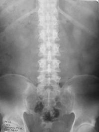
Overview urography, or to obtain a black and white image of the abdominal cavity and retroperitoneal space through x-ray machine, allows to determine the location of the kidneys, ureters and bladder. But their contours tend to be blurred due to the layering of the projections of other bodies or high lightness of the intestine. In addition, this method cannot “see” the condition of the internal cavities of the urinary organs to assess their functionality, to determine the patency of the ureters or renal pelvis.
To overcome these gaps and maximize opportunities urography, to make a real simultaneous investigation of renal excretory function, the transport role of the ureter, and cumulative values of the bladder allows the use of contrast. Due to the use of special contrasting pharmacological means method was called as a contrast urography, aka excretory urography or intravenous.
Currently in urology and Nephrology use a range of iodine-containing contrast agents, belonging to different generations and chemical groups. More modern contrasts differ the least pronounced side reactions of adults and children to their introduction. The main feature of these drugs is their ability to reflect x-rays, so the rays back into the camera and spoil the film.
The result is a sharp image in the form of white structures all spaces and cavities, where at the time of fixing the x-ray machine was a contrast substance called because of this radiopaque. It becomes possible to visualize the passage of substance at all urinary Department, to determine the structure of the renal calyx and pelvis, to evaluate the excretory function of the kidneys.
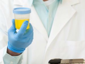
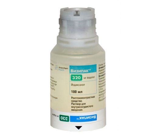
Therefore, the use of urography of the kidneys using contrast agents is more preferable for the diagnosis, exerted on the patient to radiation and chemical stress. It is better to use a nonionic contrasts, such as Urografin or Visipak better undergoing patients.
Indications for excretory urography is quite extensive both in adult and in children:
- infectious pathology of the urinary system;
- the presence of pain in the lumbar region or the abdominal cavity;
- changes in the urine;
- injury or surgery on the kidneys, ureters, bladder;
- congenital anomalies;
- urolithiasis.
Are there any contraindications to the implementation of the study
As a contrast urography is the simultaneous use of chemical substances and radiation exposure, it naturally feasible, not all categories of the population.

It is contraindicated in the following situations:
- if the patient is allergic to iodine, this means that the use of iodinated contrast is unacceptable;
- glomerulonephritis in acute stage (in this case, the excess chemical burden will worsen the condition of the glomeruli);
- acute or chronic renal failure;
- thyrotoxicosis;
- pheochromocytoma (tumor of the adrenal glands);
- pregnancy, lactation.
In these cases, preference in choosing the method of research will be given an ultrasound or MRI.
During pregnancy contrast urography is not possible
How to prepare for urography with contrast
The purpose of this research is always done individually, the doctor needs to find out the specifics of the anamnesis, presence of background diseases and allergic disposition. It is desirable to hand over the biochemical analysis of blood to exclude kidney failure. When it detects the patient has any allergies 2-3 days before urography is carried out appropriate treatment, and on the day of the study recommended the appointment of prednisolone.
Preparation for the procedure becomes for the patient too hard and burdensome. The most important point is the cleansing of the intestine and reduce pneumatization (lightness). To do this for 3-4 days before urography excluded from the diet dairy products, yeast bread and sweet baked goods, sweets, all legumes. The day of the test Breakfast is over, but you can drink unsweetened tea. In addition, it is necessary to take activated charcoal or other sorbents: 3-4 tablets 3 times a day.
Before urography should temporarily give up many Goodies
Prolonged constipation bowel cleansing it is necessary to complement mild laxatives (Duphalac) or Microlax enema. If the need arises, before urography patient put a cleansing enema directly in the hospital.
The study
General recommendation for all contrast agents is to conduct preliminary tests on the sensitivity of the patient to the administered drug. For this estimated reaction of 1 ml of iodine-containing solution for 3 minutes. In the case of the characteristic symptoms of the patient medical assistance, and urography with contrast is replaced by alternative instrumental methods.
The amount of contrast agent depends on the patient’s weight and type of drug. Thus the dose must be calculated very precisely; for example, Urografin is used in a ratio of 1 gram per 1 kg of body weight. In children, it is preferred that Visipak: 2 ml per 1 kg of weight, while the maximum number is 50 ml. If the contrast is not ink-jet, and drip, the calculated amount of substance is multiplied by 2 and diluted with 5%-th solution of glucose, taken in the same volume.
Monitoring the patient’s condition is carried out throughout the course of the procedure, which takes an average of 30 minutes to an hour. In some cases it can last an hour and a half, if, for example, the patient greatly impaired excretory function of the kidneys. Observation is necessary in order to diagnose adverse reactions. So, the patient during the procedure may be a metallic taste in the mouth or a feeling of warmth throughout the body. These conditions very rapidly and do not require correction.
Contrasting substance Visipak well-tolerated by patients
But there are also delayed complications, contrast urography, which develop after some time (usually 1 day) after the study.
They stated very rare, but still possible:
- local: hematoma at the puncture site of the vein, flebita (inflammation of the area of Vienna);
- common: hemodynamic disorders, nephropathy, renal failure.
Contrast urography is performed in the inpatient or outpatient (in the clinic). After a negative test contrast sensitivity is performed first, review the radiograph, then introduced the rest of the drug and starts the sequential fixation through images of the progress of the radiopaque medium. Thus the patient can be in supine position or to assess the mobility of the kidneys in the standing position.
Receiving a series of images taken at certain stages, your doctor may say all sorts of anatomical or functional deviations from the urinary organs, and related structures.