
Respiratory failure – a condition in which blood gas composition suffers because of violations of the processes of respiration, enabled him to normal.
10 thousand population of about 8-10 people suffer from different forms of respiratory failure. At 60-75% of patients with acute or chronic diseases of the respiratory system it was noted at least once in your life.
Causes and classification of respiratory failure
This condition can accompany most diseases of the respiratory system. But most often it occurs in diseases such as:
- inflammation of the lungs;
- cardiogenic pulmonary edema (provoked by cardiac diseases);
- respiratory distress syndrome of adults (ARDS), acute lung injury, in which there is swelling and swelling of the tissues.
Respiratory failure is:
- ventilation – in violation of the ventilation of the lungs; in this case mainly affects the respiratory tract;
- parenchymal – tissue damage the lungs themselves.
Ventilation type of pathology most often found in:
- violations on the part of the brain respiratory center;
- fatigue, weakening or loss of muscle involved in the act of respiration;
- mechanical defect of the musculoskeletal skeleton of the thorax – mostly because of congenital or acquired heart defects (most often it is kyphoscoliosis), injuries, after surgery;
- chronic obstructive diseases of the respiratory system;
- obesity.

Parenchymatous respiratory insufficiency occurs in many lung diseases is:
and so on.
Mechanisms of development
Respiratory failure is characterized by:
- excess carbon dioxide in the blood (ventilation type);
- the lack of oxygen (parenchymal type).
Speed of occurrence and development of respiratory failure is:
- sharp;
- chronic.
Acute respiratory failure is characterized by such features:
- occurs suddenly – within a few days, hours, sometimes even minutes;
- progresses rapidly;
- accompanied by disturbances from the bloodstream;
- may threaten the patient’s life, which will require intensive therapy.
Features of chronic respiratory failure:
- begins with invisible or non-special subjective discomfort symptoms;
- can develop for months and years;
- able to develop if the patient is not fully recovered from acute respiratory failure.
Important! Even if the patient suffers from chronic respiratory failure, may occur its acute form – it means that the organism has not coped with chronic respiratory failure, it is not kompensiruet.
Distinguish mild, moderate and severe degrees of respiratory failure, which confine oxygen pressure and saturation of blood: mild blood oxygen pressure is 60-79 mmHg. article, saturation is 90-94%, with an average – 40-59 mm Hg. article and 75-89%, severe – less than 40 mm Hg. article and less than 75%.
Normally, the oxygen pressure is more than 80 mm Hg. article, the saturation – more than 95%.
External respiration (i.e., the flow of oxygen through the Airways in the lungs) are supported by many links one established mechanism is:
- the Central nervous system and respiratory center;
- neuromuscular system (in particular, the structure of the chest);
- respiratory tract;
- the alveoli of the lungs.

The failure of any link will lead to respiratory failure.
CNS and respiratory center, which often lead to respiratory failure:
- overdose of drugs (including medication);
- reduced thyroid function;
- the deterioration of cerebral circulation.
Pathological condition on the part of the neuromuscular system causing respiratory failure:
- the Guillain-Barre syndrome (a condition in which the immune system reacts to its own nerve cells as foreign structures);
- botulism;
- myasthenia gravis (muscle weakness, which in turn, can evolve because of many reasons);
- disease Duchenne (characterized by degeneration of the muscles);
- congenital weakness and rapid fatigue of the respiratory muscles.
Violations of the thorax, which may develop respiratory insufficiency:
- kyphoscoliosis (curvature of the spine in two projections);
- obesity;
- state after thoracoplasties operations;
- pneumothorax (air in pleural cavity);
- hydrothorax (fluid in the pleural cavity).
Pathological conditions and diseases of the respiratory tract, due to which there is respiratory distress:
- laryngospasm (constriction of the lumen of the larynx due to the reduction of its muscles);
- swelling of the larynx;
- obstruction (obstruction) foreign body at any level of the respiratory tract;
- bronchial asthma;
- chronic obstructive disease of the respiratory system (particularly obstructive bronchitis with asthmatic component);
- cystic fibrosis (lesion of the glands of external secretion – including the airway);
- obliterative bronchiolitis (inflammation of small bronchi with subsequent obliteration).
Destruction of the alveoli leading to respiratory failure:
- different types of pneumonia;
- respiratory distress syndrome, adult;
- spadenie lung (atelectasis), which can be caused by many reasons;
- pulmonary edema of different origin;
- alveolitis (inflammation of alveoli);
- pulmonary fibrosis (massive germination of the lung parenchyma connective tissue);
- sarcoidosis (mass education in the organs peculiar nodules – including in the lungs).
Describes the causes leading to hypoxemia – decrease in the level of oxygen in the tissues.
Direct mechanisms of its origin:
- in portions of air, which breathes in man, the so-called reduced partial pressure of oxygen;
- easy poorly ventilated;
- bad gases pass between the walls of the pulmonary alveoli and the walls of blood vessels;
- venous blood is discharged into the arteries (this process is called bypass surgery”);
- pressure of oxygen in mixed venous blood decreases.
The partial pressure of oxygen in a portion of the air which breathes in man, may be reduced under the following conditions:
- within easy reach near combustion sources (uses oxygen);
- in case of inhalation of toxic gases;
- at high altitude (particularly at high altitudes) as a result of the thinning air and reduced atmospheric pressure.
Due to the fact that the lung is poorly ventilated, the alveoli increases the pressure of carbon dioxide, and this leads to a decrease in oxygen pressure in the same alveoli.
The deterioration of the passage of gases in the walls of the alveoli and blood vessels often occurs in such diseases and conditions as:
- alveolitis;
- the proliferation of connective tissue in the lungs;
- sarcoidosis;
- asbestosis (occupational disease due to work on asbestos production, causing the asbestos accumulates in the lungs);
- carcinomatosis (metastases to the lungs due to malignant tumors of any localization);
- age-related changes in the lungs;
- changing the position of the body, which may affect the amount of light.
When shunting of venous blood does not pass through the bloodstream to the lungs, and if it is, then only in those parts of the lungs where gas exchange is not observed. For this reason, venous blood gets rid of carbon dioxide, it continues to circulate in the vascular system, thereby not allowing blood to get enough oxygen. The lack of oxygen that occurs in this bypass surgery, it is very difficult to adjust oxygen therapy.
Respiratory failure due to the discharge of blood occurs in such conditions as:
- pulmonary embolism;
- shock of various origins;
- perform physical work patients with chronic respiratory diseases.
The increase in carbon dioxide develops as a result of:
- deterioration of ventilation of the lungs;
- the increase in so-called dead space (segments of the lung that do not participate in gas exchange);
- increase in carbon dioxide in the environment.
The process of lung ventilation depends on many factors that support it – from the nervous and ending with the provision of the respiratory muscles.
If increasing those areas of the lung that do not participate in gas exchange, runs the compensatory mechanisms by which the ventilation of the lung is kept at the desired level. Once these mechanisms are exhausted – there comes a deterioration in ventilation.
Increasing the amount of carbon dioxide can be observed because of its excessive revenues from the external environment, and as a result of its enhanced production by the tissues. Most often this occurs in such conditions as:
- the increase in body temperature; increase it by 1 degree leads to increased production of carbon dioxide by 10-14%;
- muscle activity is not only physiological (exercise, physical labor), but the norm is not observed (seizures, convulsions);
- enhancing parenteral nutrition – nutrients in the form of the input solutions.
Particularly parenteral nutrition affects the increased production of carbon dioxide, if increased content of carbohydrates. This mechanism is not as important for increased production of carbon dioxide, but other failures contribute to the problem.
Symptoms
Clinical symptoms shows as a lack of oxygen or excess of carbon dioxide.
The most frequent manifestations are:
- shortness of breath;
- feeling of choking;
- blue discoloration of the skin and visible mucous membranes;
- changes in the Central nervous system;
- the weakness and then the fatigue of the muscles involved in breathing.
In dyspnea, the patient makes an effort to inhale, which in the normal state is not required. The degree of dyspnea is not an indicator of lack of oxygen or excess of carbon dioxide it is difficult to conclude, as expressed respiratory insufficiency.
About the level of hypoxemia and hypercapnia (excess carbon dioxide) more clearly signal other clinical signs – changes the color of the skin, circulatory disorders and symptoms of Central nervous system.

Signs of hypoxemia:
- cyanosis – always appears in front of her. Bluish color of the skin appears typical at partial pressure of oxygen below 60 mm Hg. article and blood oxygen saturation less than 90%;
- palpitations and heart rate and increased blood pressure;
- violations of the CNS: if the oxygen pressure fell to 55 mm Hg. article, the patient is memory, if up to 30 mm Hg. article – loses consciousness;
- if respiratory distress is observed the patient for a long time, that is manifested by proliferation of bone marrow cells. This process is designed to compensate for hypoxia (bone marrow is involved in hematopoiesis, and therefore ensures normal transport of oxygen by blood cells).
The symptoms indicating an increase in the amount of carbon dioxide is the result:
- increased activity of the sympathetic division of the autonomic nervous system (the part which enhances the activity of internal organs);
- direct action of carbon dioxide on the fabric.
The most typical clinical symptoms indicating an excess of carbon dioxide is:
- violations of hemodynamics (movement of blood through the vessels);
- changes in the Central nervous system.
When excess carbon dioxide hemodynamic changes as follows:
- frequent heartbeat and pulse;
- develops vasodilation throughout the body;
- increased cardiac output of blood.
Central nervous system responds to increases in carbon dioxide levels in the following way:
- appears tremor (shaking of the trunk and limbs);
- patients suffer from insomnia if they manage to get to sleep – often Wake up in the night and the day cannot overcome the drowsiness;
- get headaches (mostly in the mornings);
- marked nausea not associated with food intake or changes in body position in space.
If the pressure of carbon dioxide increases rapidly, the patient may even fall into a coma.
The clinical manifestations can reveal the fatigue and weakness of the respiratory muscles:
- first I get short of breath (fatigue fix in that case, if the respiratory rate is 25 of the acts of inhalation and exhalation per minute);
- further, with increasing pressure of carbon dioxide breathing becomes less. If respiratory rate is less than 12 in 1 minute, this should be cause for alarm medical: this CD may indicate a possible emergency stop breathing.
Normal respiratory rate at rest is 16 to 20 acts per minute.
The body tries to maintain normal breathing that increases the muscles, which normally do not participate in the act of respiration. This is manifested by the contraction of the muscles that lead to nasal flaring, tension of the neck muscles, reduce abdominal muscles.
If the fatigue and the weakening of the respiratory muscles has reached a very extent, it begins to manifest a paradoxical breathing: during inhalation, the rib cage will shrink and fall down, exhale to expand and rise upward (in the norm is the opposite).
Diagnosis
These symptoms allow you to record the fact that respiratory failure and to assess the degree of its development. But for a more accurate assessment it is necessary to investigate blood gas composition and acid-alkaline balance.
The most important is the study of such indicators as:
- the partial pressure of oxygen;
- partial pressure of carbon dioxide;
- the pH of the blood (determination of acid-base balance);
- the level of bicarbonates (salts of carbonic acid) in arterial blood.
When the ventilation of respiratory failure, a shift in pH of blood acidic side, with the defeat of pulmonary tissue in an alkaline.
Determination of the level of bicarbonate allows to judge about the neglect of the process: if the number is more than 26 mmol per liter, this indicates a prolonged increase in the level of carbon dioxide in the blood.
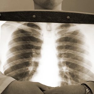
For evaluation of disorders of gas exchange is carried out x-ray light. In some cases x-ray signs may not be recorded, although the clinic says about respiratory failure.
This happens when:
- the shedding of blood (shunt);
- chronic obstructive diseases;
- bronchial asthma;
- pneumothorax;
- obesity.
On the other hand 2-sided massive radiological changes at a moderate clinic can occur when:
- massive pneumonia;
- edema of lung;
- the fluid entering the lungs;
- the pulmonary bleeding.
For investigation of breathing to understand what his team is suffering, conduct spirometry – study of external respiration. For this purpose the patient is asked to do the breath, with the specified parameters (for example, with different intensity).
Such methods help to analyze:
- how passable airway;
- what is the status of the lung tissue, its blood vessels and the respiratory muscles;
- what is the severity of respiratory failure.
While conducting such research methods is primarily determined by:
- vital capacity – the volume of air which can put the lungs at maximal inhalation;
- forced vital capacity – amount of air that the patient can exhale at a maximum speed of expiration;
- the volume of air that the patient exhales in the first second of exhalation
and other parameters.
Treatment and emergency treatment of respiratory failure
The basis of treatment of respiratory failure are:
- the elimination of the causes that provoked it;
- securing the airway;
- gaps of oxygen in the body.
Methods of elimination of causes of respiratory failure very much depend on its causes:
- if there is a respiratory infection – prescribed antibiotics;
- when respiratory failure due to pneumothorax carry out drainage of the pleural cavity;
- when obstruction due to foreign body extracted

and so on.
Chronic respiratory insufficiency insidious that its influence over the conservative methods don’t work. Recently such attempts – thanks to a lung transplant. But at the moment this method does not apply to common – the vast number of patients treated with well-established conservative methods that might facilitate to alleviate symptoms of respiratory failure, but not eliminate it.
The airway provides methods that liquefy sputum and help the patient to cough up her.
In the first place is:
- receiving bronchodilators and mucolytics;
- postural drainage (patient takes a position and begins to expectorate);
- vibratory massage of the chest.
Not even too prolonged hypoxemia can lead to death, so the gaps of oxygen in the body is extremely important.
For this purpose, use:
- oxygen therapy;
- medications to improve breathing;
- changing the position of the body;
- improvement in cardiac output.
Oxygen when oxygen therapy is delivered to the body in different ways – primarily through:
- the so-called nasal cannula (a tube with a special tip);
- simple facial mask;
- specially designed Venturi mask;
- the mask supplies bag.
Pharmaceuticals, designed to improve breathing, pick depending on what part of breathing is affected.
Despite the apparent simplicity, the method changes the position of the body (from stomach to side) can significantly improve the flow of oxygen in the blood and then into tissues. At the same time:
- under the influence of gravity redistribution of blood flow and reduction in the discharge of venous blood (shunting). The patient can lie on your stomach up to 20 hours a day;
- due to the fact that decreases compliance and a healthy lung, increasing ventilation in the affected lung.
Improvement in cardiac output is carried out using drugs that make up the blood volume.
In severe cases when other methods do not help, resort to hardware mechanical ventilation.
It is indicated for:
- disorders of consciousness, indicating a significant respiratory distress;
- fatigue of the muscles involved in the act of respiration;
- unstable hemodynamics;
- a complete stop of breathing.
Effective is inhalation of helium-oxygen mixture.
Prevention
Measures to prevent development of respiratory failure is a complex of activities, which today can be in a separate a small section of pneumology.
Prevention of respiratory failure is reduced to:
- the prevention of diseases that cause it;
- the treatment of already occurring disease that may be complicated by respiratory failure.
It is very important to prevent the development of chronic respiratory failure that is difficult to correct.
Forecast
Even non-durable hypoxemia can lead to death. Rapid diagnostic and therapeutic measures in acute respiratory failure help to eliminate it without consequences for the organism. Action in chronic respiratory failure help to relieve symptoms but not cure it.


I am curious to find out what blog system you’re using? I’m having some minor security problems with my latest website and I would like to find something more safe. Do you have any recommendations?