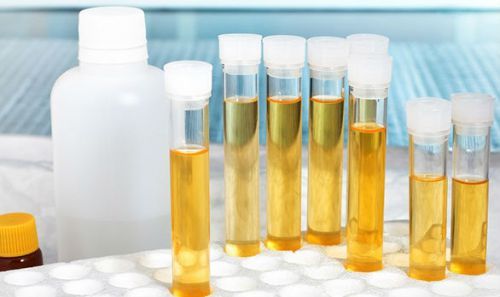
Effective method for diagnosing, monitoring the course of disease is urine microscopy. Urinalysis is also done in the case of screening. In the clinical urine sample is evaluated physico-chemical characteristics of urine microscopy and urine sediment.
Preparation to gather material for the analysis of urine is preceded by the holding of personal hygiene, to prevent bacteria. Taken the morning urine obtained after the spill.
Period of time designated for delivery of the material in the laboratory, the minimum cut. Collect urine in a sterile container and then placed in a vial with preservative.
That affects the result
The laboratory result will not be reliable:
- For violation of instructions for delivery of the material (errors in hygiene procedures, the collection of material during menstruation).
- If the urine is stored for several hours before you get to the lab.
- The presence of excess amounts of fluid intake before testing, failure to comply with dietary requirements.
- If eve was performed by parenteral administration of saline, glucose solutions, contrast agents.
- The presence of a strong physical stress, nervous disorders.
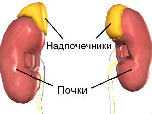
- In pregnant women.
- If the urethra is traumatized by the catheter or with a smear from the urethra.
- If there is contamination hemorrhoidal blood.
- When taking medicine, changing individual parameters.
Collection of urine

The purity of the urine depends on the accuracy of the study
Physical characteristics of urine
Microscopic examination of the physical indicators of urine includes: color, clarity, odor, density of urine.
Color
Urine contains pigments that give it a yellow color, differing in the degree of saturation.
Color can change:
- disease;
- foods;
- medicines.
Diseases accompanied by jaundice, giving the urine a dark shade of yellow. When blood appears reddish. If the food there is an excessive amount of carrots, urine will become orange color, beet red.
Transparency and smell
Normal urine is transparent, after standing becomes cloudy: salt precipitate. The gain of the smell occurs when you access the urine of air. Diabetes feel fruit shade. Foods change the smell of onions, garlic, horseradish. Medical drugs have this same ability.
Density
At different times of the day the density of urine varies. When filtering blood, the kidneys secrete primary urine. When the opposite happens suction discharge, she re-enters the bloodstream. The secondary urine is formed with a higher concentration.
Chemical study
pH is the ratio of hydrogen ions and hydroxide ions. If their number is equal, a neutral solution. In case of imbalance, the solution will be acidic or alkaline. Normal urine is characterized by a slightly acid reaction. Shifts in the reaction of urine in one direction or another dangerous formation of stones.
A shift to the acid side occurs:
- if used an excessive amount of protein;
- at the phenomena of fever;
- if the body is subjected to heavy loads;
- with all the types of fasting;
- if the body is affected by diabetes.
The alkaline side:
- in case of refusal from animal proteins;
- frequent vomiting;
- in the pathology of urinary system.
Protein
With normal rates of protein in the urine is missing, rather, is in such quantity that it is possible to detect, using special research methods that would be impractical.

Protein in the urine indicates the presence of the disease
To determine the presence of protein, the urine must be mixed with the desired reagent. The presence of protein is determined by the presence of a reaction: if a reaction occurred – a protein present in the urine. The presence of protein in the urine is called proteinuria.
The emergence of functional proteinuria occurs in the following cases:
- if the body is exposed to physical stress;
- in the case of nervous disorders;
- with the prevalence of protein;
- in febrile phenomena.
Glucose
Normal glucose in urine should not be. Sometimes microscopic analysis of short-term notes the emergence in the treatment with glucose or other increased sugar uptake by the body. Most often, the glucose found in diabetes.
The presence of glucose in urine is called glucosuria. There are methods for the detection of glycosuria. For this purpose, use a test system strips for applying droplets of urine to monitor a color change. Comparing the change of the rating scale, it is possible to give an opinion on the amount of glucose in the urine.
Ketone bodies
Detection of ketone bodies in the urine is considered a deviation from the norm, the reason for such situation is the occurrence of disturbances in the metabolism of fats and carbohydrates. If the body has to get energy from fat reserves when fat breakdown ketone bodies are formed, which extend from the body with urine. In this case, there is ketonwrïya.
The emergence of ketonuria contribute to:
- strong physical exertion;
- nervous disorders;
- illness accompanied by fever;
- the diet excludes carbohydrates;
- recurring vomiting.
Ketonwrïya is determined by using test strips with applied droplets of urine like the detection of glycosuria.
Bile pigments
Bilirubin, urobilin, bile acids under normal performance should not be present in the urine. If they were, then, in the blood level of bilirubin is elevated. The place has the appearance of bilirubin, which is characteristic of hepatitis and obstructive jaundice.
Microscopy of urinary sediment
To obtain the precipitation of urine is needed about 2 hours. The elements found in the sediment, divided into organized and unorganized. The first are of organic origin, the second is inorganic. Microscopy of urine, there are permissible limits of the presence of the elements of the urinary sediment. If these figures are exceeded, one can assume the development of various pathologies.
Hemoglobin
In the investigation of urine hemoglobin is found in urine in heavy infections of the body. Hemoglobin appears in the urine during the disintegration of red blood cells. The reasons for this condition are often of diseases caused by various infections.
To provoke the emergence of hemoglobin in the urine can:
- re-hypothermia;
- physical activity;
- severe poisoning;
- exercise.
In such cases, the urine changes colour, becoming red-brown, in the lumbar region the pain is felt. Often the urine is saturated with hemoglobin in case of unsuccessful blood transfusions. This occurs when the incompatibility between blood donor and the receiving patient.
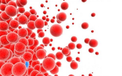
Erythrocytes
There are 2 types of erythrocytes present in the urine of intact and leached. The last does not contain hemoglobin. Leached erythrocytes indicate pathologies of the kidney, unaltered – about violations in the urinary system.
The norm of erythrocytes for women – 3 men – 1. If the figures differ, there is hematuria, which is of two types: microscopic hematuria in which the color of the urine remains unchanged, and gross hematuria (urine color changes because of the presence of red blood cells).
Hematuria (increased presence of red blood cells) appears in the following cases:
- at pathologies and injuries of the kidneys;
- in various forms of diathesis;
- if kidney stones are present;
- for cancer of the urinary system;
- when injected into the body of toxic substances.
Leukocytes
Plenty of leukocytes in urine leads to its dimness. For women a normal leukocyte count in urine, considered 0-5, 0-3. Increase of indicators indicates the presence of pus in urine that signals the existence of inflammatory processes.
To determine where the inflammatory lesion, is glass test of urine, in this case trehstakannaya. The first glass contains samples of the initial pyuria, indicating the presence of urethritis, prostatitis. The number of leukocytes in 3-Cup determines the final pyuria, which often indicates the disease cystitis.
Excess of leukocytes in three cups indicates diseases of urinary system.
Epithelium
The presence in the urine is considered normal. Increase the valid values of the squamous epithelium confirms severe inflammatory pathology. Polymorphic epithelium above the norm occurs in cancer, the presence of stones, defeat different kinds of intoxications.
Cylinders
Found in different kidney diseases. The presence of hyaline cylinders allowed only in the unit quantity as causes of are physical exertion. Other types of cylinders in norm should not be available. Their presence in urine is called cylindruria.
Reasons cylindruria are:
- kidney disease;
- pathology of blood circulation;
- diseases accompanied by fever;
- exposure to various infections.
Unorganized sediment
Contained in urine of salt should not exceed 30-40 mg. If the work of the urinary system is broken, then the salts accumulate. In acidic urine, consisting of salt urates appear. Alkaline urine will accumulate ammonium urate, amorphous phosphate. Oxalates are present in both types of urine.
Bacteria in the urine
Normal bacteria in urine are not present. Clinical analysis establishes the presence of bacteria. To identify these species, conducted bacteriological seeding. If found isolated bacteria, raises the diagnosis of bacteriuria. Cause infection of the urinary system.
The technique of quantitative studies of urine
When you use these methods receive more detailed information about elements in the urinary sediment provides assistance in diagnosing latent pyuria. Methodology quantitative research with microscopy gives the possibility to count elements at a specific time in a specific volume.
It should be noted that in children if there are indications studied urine samples as well as in adults. Interpreting the results takes into account age-related norms. Often for the study of the urinary sediment using the method of Nechiporenko, in which manual digital processing is replaced by fast and accurate calculation for a given algorithm implementation.
The sequence of the microscopy of urine on Nechiporenko:
- Prepared urine transferred into a centrifuge tube.
- Urine centrifuge is processed at a speed of 1500-2000 rpm in the course of quarter of an hour.
- The urine separated from the precipitate to a test tube 1 ml of urine sediment.
- Does the slide-plate, and his cell is filled with a drop of urine sediment.
- Slide the tablet is placed on the microscope stage, and his help counts the number of formed elements.
- Using a special formula, set their number in 1 ml of urine.
After receiving the results for the diagnostic output using the Atlas of microscopy of the urinary sediment.