In the diversity of research techniques an x-ray of the kidneys is one of the first places in the diagnosis of diseases of the urinary system. Radiography of the kidneys provides the opportunity to study in detail its structure, pathological changes in the body.
This technique appeared a long time ago and still has not lost its relevance, thanks to the valuable information that it is able to give.
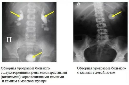
Depending on what pathology is suspected by the doctor, he recommends that one of the varieties of x-rays of the kidneys. The most basic method is a panoramic picture that shows a picture of the location, size, and configuration of the kidneys.
The rays of the x-ray unit aimed at abdominal part of the body, so before the procedure, the bowel must be cleansed. Contrast material is not required.
Varieties of x-ray
The other mode is computed tomography. It is a methodology which gives the possibility of using x-rays to obtain cross-sectional image of the body. No preparatory procedures are not required. It is important to know that CT is a rather expensive method of diagnosis. Perhaps this is its only drawback. The rest of it is much more efficient than regular x-rays. CT scan reveals the tumor much more accurately and faster.
Another type is the x-ray of the kidneys with contrast. Spend it in different ways. It is administered only in the hospital but not outpatient.
Radiograph with contrast
It is this kind of determining the cause of kidney disease is very serious. It is carried out only on very strict conditions. A kidney x-ray with contrast is divided into subspecies on the basis of the contrast agent.
Release:
- intravenous urography;
- direct pielografia.
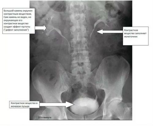
Intravenous urography form requires the input of contrast (iodine-containing substance) into a vein. Injected with a current of blood enters the kidneys, later in the ureters and bladder. While doing a series of shots, which shows how the contrast material moves through the system the urinary tract. This produces an x-ray of the bladder, which can be seen in the pictures, as well as of the ureter.
The contrast is displayed naturally. In the case of disorders of the kidneys (based on analyses), a contrast agent is injected very slowly, the dosage, while monitoring the urinary system. This subspecies is called intravenous urography.
Contraindications to this kind of research will be:
- intolerance to iodine;
- child bearing and lactation;
- heart failure;
- reduced blood clotting;
- kidney disorder;
- increased activity of thyroid hormones.
There is also a method such as meccinna cystography, in which you can get an x-ray of the bladder. In this case, the movement of a contrast agent evaluated during dewrinkle.
Direct pielografia
The advantage of this survey is that it provides a better look at the Cup – pelvis-plating system. Among them there are also special subspecies. The first of these is antegrade x-ray contrast solution is injected directly into the kidney. Doing it via catheter or injection. The contrast agent is displayed at the current urine.
But during retrograde pyelography, the contrast carried out through the catheter, but against the normal flow of urine. The contrast gets into the urethra, bladder, ureters and kidneys. Do not use this approach in the presence of inflammation and hematuria.
Angiography
This radiopaque study aimed at the visualization and study of blood vessels that nourish the kidney and the expected growth. Contrast is provided in the vessel and gives color. The first subspecies is a common angiography, a catheter is put through the femoral into the aorta, the end portion of it is put in place branches from the aorta to the renal vessels.
The second subspecies is called selective angiography. In this case, the catheter is placed directly into the artery of the kidney. But with selective venography professionals need to image the venous system of the kidney. To this end, through the inferior Vena cava, the catheter is introduced directly to these vessels.
Indications for x-ray
Indications for radiological research is extensive.
The main ones are:
- pain in the lumbar region with the exclusion of neurological disease;
- hematuria and mucus in the urine;
- odor of urine (smell of fish);
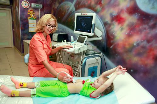
- pathology ultrasound of the kidneys and bladder;
- a disturbance in the General analysis of blood and urine;
- pain when urination;
- suspicion of the presence of stones.
What diseases can be found
Radiographic kidney give the opportunity to detect a number of diseases, including:
- kidney stones location of stones;
- tumors;
- polycystic;
- cysts of the kidneys;
- the state after grinding of stones;
- nephroptosis;
- dropsy of the kidney;
- the inflammatory process the layers of fat tissue;
- inflammation of the kidneys;
- injury of the urinary tract and blockage at different levels;
- deviation in the development of the organs of mochevyvodjashie;
- tuberculosis;
- pyelonephritis, and glomerulonephritis;
- renal hypertension.
As you can see, the list is impressive.
How to prepare yourself for the procedure?
Preparing for x-rays of the kidneys is very serious matter. Three days before the intended examination is necessary to establish a diet. It consists in the exception of gas-forming food products. It is sweets, cakes, beans, cabbage, black bread, milk, soda, fruit.
If the patient has problems with the chair, then three days need to start taking laxative products (lactulose, sentex, magnesia). Before the procedure the last time you can eat at six o’clock. In the morning you must do an enema.
The stages of x-ray
When x-rays of the kidneys, the survey depends on the type. If it’s plain radiography, the patient is free from clothes upper body, then laid on the couch, he placed a cassette tape. Then take a series of shots, if necessary, repeat it in a vertical position.
If doing a contrast x-ray of the kidneys, the patient carry out a tolerance test of iodine. In the shoulder under the skin, administered one ml of contrast, and to control in the other hand also subcutaneously injected the same amount of saline. After twenty minutes, inspect the result, if there is hyperemia of more than 3.5 mm in diameter, we are talking about allergies. Then the examination is canceled. But if the skin is clean, a contrast agent is used.
A sick person should know that during the procedure might be discomfort (redness of the face, pain, burning). But if he feels itchy, watery eyes, difficulty breathing, the survey should immediately finish.
Pielografia also suggests a test for an allergic reaction to iodine. If no allergies, then the catheter, which introduces a contrast. Sensation with the introduction of unpleasant. In the case of the antegrade pielografia the patient is given a mild anesthesia, place the catheter to numb. Then enter it in the kidney.
Urography and pyelography a list of suggested pictures that are shot in a horizontal and vertical position.
Examination in children
This method of diagnosis has its own principles in children. Is a kidney x-ray purely on the testimony, if the ultrasound its possibilities have been exhausted. Children are given a light anesthesia. Examiner only in the presence of two doctors. If there is a need for repeated procedures, then implement them across large gaps. The dose of the contrast calculated on the basis of body weight of the baby.
A small child usually needs fixing, this is the job dad and mom under cover aprons of lead. Children also recommends carminative bloating for 3 days before the procedure, and the kid, under one year, forbidden to eat beans, sugary drinks, fruits. 7 hours before excretory urography do not drink. Feedback from parents about this method of diagnosis have a positive color.
Parents must take the procedure a bottle with milk or porridge. This is necessary to ensure that in the presence of high gas content of the intestines, by means of filling the stomach to give the opportunity to carry out the survey. Also nice to have a toy and a pacifier.
How to interpret the result?
You need to understand that buds are visible not all people, in 40% of patients, they are not visualized. The others they represent in the picture of shadow education, which is from the twelfth vertebra of the chest on the second vertebra of the lower back (left kidney) or from 1 to 3 lumbar vertebra. The ureters, prostate gland is not visualized, the bladder can only be seen when it is filling. The x-rays also you can see the position of the adrenal glands.
Retrograde pyelography and excretory urography are distinguishable pelvis of the kidney, in terms of size they are comparable with the shadow of the kidneys on survey radiographs. Pelvis in the form of capsules and dendritic is a normal form in which no need to worry. Using contrast visible ureters (size, shape, place of origin and entry into the bladder). In the absence of pathological changes it is not enlarged, no stones. In addition, the doctor looks for a leak of contrast, which happens when the tears in the urinary tract. When the contrast reaches the bladder, it is possible to evaluate its configuration (normal pear shape), shaped smooth.
As for kidney stones, even in their presence, they are not always visible. In the presence of gases in the intestine can not see them. Also not visible stones, consisting of oxalic acid, phosphates, and the overlay image of the stone on the vertebrae.