The diagnostic method is determined only by the doctor on the basis of a conversation with the patient, his external examination, examination of tests, medical history and other information. In complex cases, several research methods are used.
The complex clinical picture of some pathologies of the human excretory organs requires the passage of a whole range of studies on different devices. Due to the different information content, each of the devices gives out its information. Therefore, you need to choose what will give the maximum data for determining the diagnosis.
It is not entirely correct to raise the question of the choice between CT, MRI and ultrasound. Such studies perform several different tasks. The information of each of them is added to the overall picture, making up for the missing parts. As an initial examination of the kidneys, the doctor often prescribes an ultrasound. If there is not enough information, sometimes an MRI or CT scan is performed. Tomographs are used in difficult situations when other methods fail.
In some cases, if there are contraindications, only ultrasound is prescribed to detect the disease. For example, in a first trimester of pregnancy, a doctor will never prescribe a kidney tomography. Such a procedure can harm the health of the unborn child.

In any case, in any research, it is better to fully trust the recommendations of specialists.
The most important paired organ of a person with the role of a natural filter is the kidneys. Its task is to clean all body fluids from unnecessary substances and remove them. In addition, it performs many other functions. This organ is susceptible to various diseases, their symptoms often coincide, and sometimes are completely absent.
The disease sneaks up imperceptibly. You can find out about the condition of the kidneys if you pass the necessary tests and undergo the necessary examinations on modern diagnostic devices. The most common of these are computed tomography (CT), magnetic resonance imaging (MRI) and ultrasound (ultrasound). Each has its own advantages and disadvantages. Which is better to choose? Let’s try to figure this out.
CT scan
The principle of operation of the device is based on the properties of x-rays. It has some similarities with the operation of the x-ray machine. The radiation source of the device emits rays that pass through the patient’s body and fall on special detectors.
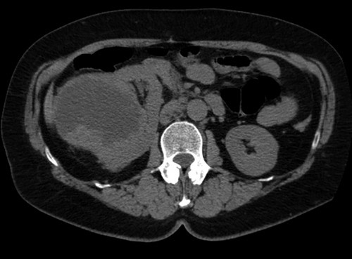
Before the procedure, to increase information content, a special contrast agent is introduced into human tissue . The tomograph takes a large number of x-rays, after which the information is processed and displayed on the monitor as an image.
Such a study allows you to consider:
- The position and shape of the kidney and adrenal gland, pathology in them.
- Tumors, their nature, hemorrhages and necrosis.
- Renal cysts.
- The development of obstacles to the outflow of urine from the urinary canals, abnormal accumulation of fluid.
Magnetic resonance imaging
By informativeness, the method is considered one of the best and safe. The device registers changes in electromagnetic fields as they pass through different tissues of the body. The basis of the device is a large ring-shaped electromagnet, into which the patient is placed.
Scanning is performed using a strong magnetic field. The received information is transmitted to a computer, processed, displayed on a monitor. The doctor examines the volumetric image of the investigated area, makes a conclusion.
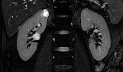
An examination of the kidneys on MRI allows you to find pathologies, as well as determine:
- Their position and parameters.
- Shape.
- Cortical and medulla.
- The condition of the ureters.
- Cup-and-pelvis system.
- Arteries, veins and nerve trunks of an organ.
- Adrenal.
- Fatty tissue.
- The surrounding tissue.
Ultrasonography
This is one of the most common hardware diagnostic methods. It captures the reflection of ultrasonic waves passing through the tissues of an organ. The extracted information is processed and displayed on the screen or printed on paper. As a result, a two-dimensional image is formed.

The diagnostic process is very simple. The doctor applies a sensor to the body of the lying patient and analyzes the image received on the screen. It is possible to print a picture.
Such a study of the kidneys allows you to establish:
- Structure, organ parameters.
- Jade, pyelonephritis.
- Vascular disorders.
- Cysts
- Tumors
- The presence of stones, sand, as well as their effect on the state of the organ.
- Anomalies of development.
- The condition of the ureters.
What do they have in common
- The listed diagnostic methods are quite safe, informative, allow you to draw certain conclusions and conclusions on the state of the investigated body.
- Each of them performs a deep scan, the result of which is displayed on the screen or paper.
- The result is obtained without internal intervention in the body.
- The procedures are painless for patients, unlike a biopsy or surgery.
Comparison of diagnostic methods
Compare the presented research methods according to several criteria:
- Availability.
- Physical phenomena used in the apparatus.
- Informational content.
- Security.
- The presence of contraindications.
- The duration of the procedure.
- Cost.
One of the simplest, affordable, and most common methods for examining kidneys is ultrasound. This device is available in almost every medical facility. There are portable models of the device. In this design, it can be delivered directly to the patient’s bed. MRI and CT are less accessible, such devices are more complex and bulky.
All these methods use different physical phenomena in their work : in CT they are X-rays, in MRI they are an electromagnetic field, in ultrasound they are ultrasound. All of them act differently on human cells, causing different consequences.
The most complete and accurate description of pathologies is given by MRI. Among the listed methods, it is the most sensitive. Further as descending are CT and ultrasound. The latter method may not reveal violations of the structure of the kidneys, but, at the same time, their functionality and performance will be reduced.
The safest method is ultrasound. It does not have any negative effect on body tissues; it can be used for a patient of any age. Computed tomography has several limitations due to x-rays.
With the exception of ultrasound, for other methods there are contraindications for use. CT is forbidden to do with an allergy to iodine, children, during pregnancy.
MRI has a much larger list of prohibitions:
- Metal bodies, implants inside.
- Pacemakers.
- The first, third trimesters of pregnancy.
- Claustrophobia.
- Tattoos with metal paints.
- Epilepsy.
- Exceeding the patient’s weight is greater than the permissible parameters.
The shortest study time on a computed tomography scanner. To reduce the time of the negative effect of x-rays on the tissue, the designers reduced the procedure to the maximum. The duration of the procedure for MRI reaches one hour.
All of the listed devices belong to expensive equipment. The cost of the study takes into account the price of the device, maintenance and consumables. The cheapest will be an ultrasound scan, the most expensive is an MRI scan.