Ultrasound of the kidneys belongs to the category of the safest surveys, and this procedure has a high efficiency, and is used for detection of various pathologies of the paired organ.
If you suspect kidney disease or kidney stones, the urologist needs to send the patient for ultrasound diagnostics.
Using this method it is possible to determine abnormalities in the size of the body, stones, sand, tumor formation, brushes of various sizes. With early diagnosis of cancer, you can start a quick and timely treatment that will allow you to preserve human life.
The procedure is performed using ultrasound and transducer, which is applied to a special gel. Manipulation is completely painless, therefore, does not require the use of drugs and injection of a contrast agent. The survey does not require complex skills, but must only ultrasound diagnostician. The main condition that must be met is proper preparation prior to the manipulation.
The advantages of the method
As mentioned above, ultrasound is a safe and informative procedure. This method does not bring any discomfort to the patient, at the time of its execution there is no pain and the risk of complications. Because of this, it can be used even by infants, so as to collect urine for the primary analysis in infants is very problematic.
Before ultrasound of the kidneys need to visit the urologist or the therapist because the professional will tell you how to pass the training, will explain the results and on this basis prescribe appropriate treatment. Manipulation is performed by a doctor-wistom in specially equipped rooms.

Renal ultrasound is a completely safe procedure, so it can be done for all categories of patients
The technique of the procedure is that through the tissues of the human body pass ultrasound waves, which are tuned to a specific frequency. They have the ability to bounce off of all tissue structures and organs, resulting in a formed pattern, which is displayed on the computer screen.
The advantages of ultrasonic diagnostics:
- security;
- informative;
- quick results;
- requires no pain medication;
- efficiency.
Full ultrasound safety is confirmed by the fact that it can be done even infants and expectant mommies. Some patients think that the device creates the harmful radiation, but in fact it is not. Accordingly, manipulations of this kind can be performed as needed, namely for observation of the pathological state in the dynamics. No negative impact on organs and systems is not observed.
In the process of diagnosis can detect changes in the size, education or change of position of pairs on, these parameters allow to estimate the information content of the method. Renal ultrasound with Doppler (Doppler ultrasound) is necessary in order to check the status and patency of renal vessels.
In ultrasonic inspection, it is sufficient 10 to 15 minutes to detect abnormalities. All diagnostics can be performed quickly as it does not require specific skills and lengthy preparation prior to manipulation. During the study, the doctor immediately informs the nurse that she puts them in the form of research. The result is given to the patient and he referred to his attending physician for interpretation.

Renal ultrasound with Doppler allows you to determine the current pair of vessels on
The procedure is completely painless, so the need for the introduction of painkillers disappears. The study on the stomach of a patient sensor, and perhaps this will be painful impulses, but this is not a survey, but is a symptom of the disease. In comparison with other tests (CT, MRI), ultrasound is not very expensive method of diagnosis.
In some health care institutions to do a renal ultrasound free of charge, by referral from a doctor or coupons, but to choose a specialist in ultrasound diagnostics is not. Despite the fact that the use of ultrasound is completely safe for the human body, it is recommended to do only when necessary, and prescription.
High quality and correct result of the analysis will depend on the preparation of the patient for the procedure, the skill of the doctor and equipment.
The indications for the procedure
There is a list of readings, after which it is recommended to undergo ultrasound examination of the kidneys. Most often this procedure is used in cases of suspected urolithiasis and to verify the pathologic process in dynamics.
Ultrasound examination of the kidneys shown in the following cases:
- painful backache in the lumbar region;
- abnormalities in the clinical analysis of urine;
- injury;
- renal colic;
- pathology of the endocrine system;
- kidney disease;
- swelling.
Pain in the lumbar region, which are regular – this is a direct indication for the ultrasound. The pain may worsen in the process of walking, and usually is the main symptom of a pathological condition fresh body. Abnormalities in the indicators of urine is the presence in it of red blood cells, a color change, the presence of protein inclusions and salts, and possible sediment and turbidity.
In this case, the doctor prescribes additional examinations to determine the exact causes of deviations. If a person was informed of the injuries to the lumbar or abdominal region (falling from height, car accident), this can lead to kidney failure, so with the help of ultrasound can evaluate their performance.

Diseases of the endocrine system are a direct indication for carrying out of ultrasonic diagnostics
Renal colic is a signal that a failure has occurred in the work of the genitourinary system. The patient experiences severe painful backache in the lumbar region, radiating to the stomach and leg. The intensity of the pain can be so strong that sometimes without the use of drugs and ambulances are not enough. Often, the colic is accompanied by nausea, followed by vomiting, upset stomach and pain during urination.
Sometimes doctors confuse the symptoms and may suggest appendicitis. It is through renal ultrasound in adults and children can be a final diagnosis.Pathology of the endocrine system often produce complications in the kidneys. Such patients are recommended to undergo this analysis in order to assess the gravity of the situation.
In the presence of suspected kidney disease, people undergoing ultrasound examination to confirm or refute the diagnosis. If the disease is confirmed, the doctor will prescribe a second ultrasound to assess the development of the pathological process dynamics. Swelling of tissues indicates that kidney disorder and the fluid retained in the body, so you need to seek help from a doctor.
Preparations for the diagnosis
Preparation for ultrasound of the kidneys consists of several stages, it requires no special skills and is quite simple to implement. You must adhere to specific diets to use drugs and to drink the required amount of fluid before the procedure.
Before the renal ultrasound apparatus the patient should drink a certain amount of fluid
Before ultrasound of the kidneys of children and adults, observe the following stages of preparation:
- Many people have a tendency to increased gas-formation, to avoid errors in the diagnosis, a few days before the procedure should start taking these drugs: coal tablets, Drops, Filtrum.
- Three days before the examination from the diet should exclude the following products: soda, white bread, dairy products, cabbage, beans, soy, fresh vegetables, beer. Your doctor may prescribe a cleansing enema which should be done the evening before and the morning of analysis.
- Before the procedure it is recommended to drink plenty of fluids (400-600 ml) for filling the bladder.
One should take a normal towel, or purchase paper, to remove the residues of ultrasound gel from the skin, as most professionals is not available in the office of wipes for cleansing. This bonus is only present in private clinics.
Methods of ultrasonic diagnostics
Despite the wide popularity of this method of diagnosis, some people, the question arises: “How do renal ultrasound?”. Specific skills to perform this procedure is not required, training does not occupy much difficulty. The patient should lie down on the couch in position on the abdomen, on the side or the back.
Pregnant women often find nephroptosis. For its diagnosis it is necessary to adopt a standing position, the patient must make the maximum inhale and exhale. In order to understand how to conduct ultrasound of the kidneys, you need to understand how it works ultrasound. On the skin of the patient applied the gel for ultrasound, on top of which the doctor holds the transducer the ultrasonic waves are not audible to the human ear.
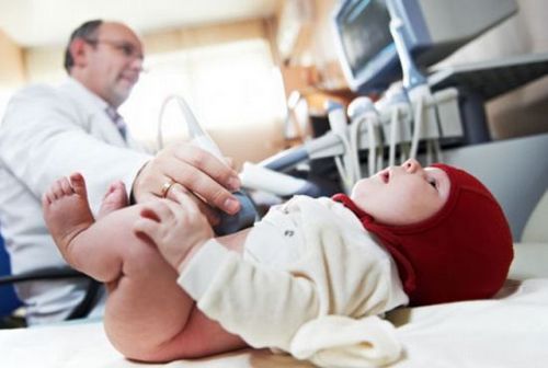
Renal ultrasound is the only method of diagnosing diseases of the genitourinary system newborn
With the help of gel improves the movement and contact of the sensor with the skin. Ultrasound waves directed at a pair on, there is a reflection effect and computer monitor displays an image. It is formed due to the fact that the waves pass through the body at different speeds. For example, using bone ultrasound passes with greater velocity than through the air.
On the screen formed a pattern, which clearly shows the kidneys as well as possible tumors if they are present, the doctor needs to do their measurements and conclusions based on what he saw. In order to assess the blood flow in the vessels fresh body, conduct ultrasound of the kidneys with the DRC. The DRC or color Doppler imaging shows on the monitor the classic image as the ultrasound, but study area is painted in a certain color, it’s fairly clear view of the flow of blood through the vessels.
Ultrasound in babies
Children’s renal ultrasound is sometimes the only diagnostic method that can be used for small children. The use of ultrasound include in a comprehensive examination of the child’s body in cases of suspected anomalies or diseases (pancreatitis, splenomegaly).
Some mothers believe that their baby is still very small and not worth it to expose the ultrasonic treatment, considering that it can cause harm. But, in fact, ultrasound is one of the safest methods of examination, since there is no radioactive radiation, and in the place where the sensor is in contact with the skin does not undergo any adverse changes.
The child’s body is not working at full strength, and therefore, before analysing children are encouraged to drink quite a different volume of fluid, in accordance with their age. Children aged one and two should drink 100 ml, from 3 to 7 years – 200 ml, Junior high school students – 300 ml and teenagers – 400 ml.