Radioisotope methods of study are very popular. They allow us to estimate the functional state of organs. To understand what it is renography kidneys, you need to define the methodology and evaluation of results.
The method
Intravenous injection of the radioisotope substance leads to its accumulation in renal tissue. The method is based on the ability of the kidneys extract from the blood hippuran and take him with urine.
On his identification with scintillating sensors, which are installed above the kidneys. The result is shown in the form of two curves.
Using this method, you can:
- to evaluate the evacuation function of the tubules;
- to determine the status of renal blood flow;
- to detect reflux from the bladder into the ureter;
- assess the condition of the tissues of the body;
- assess renal function after transplantation.

The method allows to evaluate separately the status of the right and left kidney.
Testimony
Research is indicated for assessment of the functional state of renal glomeruli in patients with the following pathologies:
- arterial hypertension;
- of renal disease of any origin;
- diabetes mellitus;
- swelling of unknown origin;
- causeless temperature increase;
- diseases of the urinary system;
- status after organ transplantation;
- any operation on the kidney.

Diabetes is one of the contraindications to the study
As a method of primary diagnosis is used when pathologies:
- kidney stones;
- amyloidosis;
- chronic glomerulonephritis;
- pathology of renal arteries;
- hydronephrosis of the kidneys;
- renal failure.
If the patient has renal anomalies– the wrong location, nephroptosis, doubling of kidneys, horseshoe, the survey results can be distorted by 50%.
Contraindications
The procedure is safe, gives large radiation loads. Introduced labeled substance is rapidly excreted in the urine. The procedure is not complications. The only condition in which is not recommended – pregnancy. Study safe even in children.
Preparation for the examination
The method is simple and versatile, special training is not required. There are certain conditions under which it is better to carry out. The patient needs to be fed. Before the study, it is recommended to drink a glass of water without gas.
Those who take diuretics the day before the study cancel. They have a diuretic effect and can distort the analysis.
The training of children has some features. They are mandatory, to prevent the thyroid gland are labeled with radioactive iodine, give the solution in drops. For these purposes, you can use Lugol’s iodine 3 drops daily. Young children in the form of games is applied to the skin iodine mesh.
The methodology of the
The survey is conducted in the sitting position. With the exception of patients who are unable to sit and children. Not conducted a study of people in a condition of alcoholic or narcotic intoxication with metal objects. The main condition for obtaining accurate results – fixed position of the subject.
In the projection of the kidneys on the back is equipped with sensors that registered the radioactive rays of labeled iodine. The location is determined based on the data of topographic anatomy. In obese patients, people with kidney migrating to Refine the localization perform x-ray picture.
Intravenous – hippuran and beginning to register its appearance in the kidneys. The amount of the drug is calculated individually depending on the age and weight of the patient. Gradual increase in the concentration, excretion of the drug is recorded in the form of the curve for each individual kidney. The survey takes 20-25 minutes.
Evaluation of results
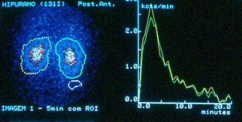
The entry curve is divided into 3 parts:
- Vascular part reflects the time of occurrence of hippuran in the vessels of the parenchyma.
- Secretion (tubular) secretion of substances from the blood by tubular epithelium.
- Excretory component excretion of the labeled isotope from the kidney.
Curve by which to assess the results of the diagnosis
The curve is placed between the two axles. On the abscissa are located the time stamp, in minutes, on the y-axis is the concentration in percent.
The norm recognized by the following characteristics:
- the time at which appears the maximum concentration of the substance 3-4 minutes;
- the time during which the curve will be half – half-life, 10-12 minutes;
- the difference between the two kidneys in these two indicators in norm does not exceed 20%, such renogram called symmetric.
Deviations from the norm this graph allows you to determine at what stage the kidney failure, but it has no specificity relative to a specific disease.
Glomerulonephritis is an autoimmune kidney disease affects both organs simultaneously, in contrast to pyelonephritis. Therefore, on the curve of the change to be visible on the right and left result. Changes for amyloidosis is relatively similar to the glomerulonephritis, bilateral. This will slow the excretion of hippuran, but kept the period of maximum concentration of the substance. If the disease progresses, the amplitude of the curves decreases, they become flat.
In hypertension caused by lesions of the renal arteries, left and right curves will be asymmetrical. On the affected side the time during which the concentration reaches a maximum, will be increased. But at the same time retain the ability to concentrate and to withdraw the same speed for both sides.
The method allows to determine renal failure before the onset of clinical symptoms. The curve will be significantly increased the time of blood purification from the radioisotope material.
Abnormal curves
Some types of research results are specific and have the names:
- Nonfunctioning type – a vascular segment and the gradual decline of the line.
- Ezoterichesky curve line rises to a certain level and throughout the study is parallel to the x-axis, which is characteristic of chronic renal failure.
- The obstructive type occurs when there are barriers to the outflow of urine – stone, tumor, adhesions. A vascular and secretory segment, but no excretion of substances (the curve gradually rises up).
- Parenchymal – reduced the height of the line, slowing down the rest of the factors in glomerulonephritis.
Renography – effective study of the kidneys, which has minimal impact on the human body
Despite the fact that renography uses a radioactive drug, this study has a very low radiation exposure, unlike x-ray or CT method. Therefore, it can be done several times. For some cases requires the estimation of the treatment, degree of recovery of renal function.
After organ transplantation, there is a need in the evaluation of the new kidney, she is functionally active and has not been damaged. This method in conjunction with ultrasound, will be indispensable.